
Symmetry analysis of the pelvic ring
Background
Patient specific implants based on additive manufacturing principles hold great promise in CMF applications, where anatomical fidelity is paramount. As the imaging to printing workflows improve, one of the major remaining hurdles is the development of bioinks that are osteoinductive, while at the same time having a realistic path through regulatory approval.
By avoiding complex material developments and following a "less is more" approach, we believe a novel material with clinical approval can be obtained more rapidly. This project investigated the printability of three clinically approved natural materials based on hyaluronic acid, fibrin and collagen. Further addition of Polyphosphate nanoparticles and dexamethasone releasing microparticles was investigated, with osteogenic differentiation the measured outcome. The potential to improve the gels osteoinductive properties by the addition of simple osteoinductive molecules that are already clinically approved would have a less challenging approval process.
Goal
To demonstrate the feasibility of symmetry analysis of the pelvic ring using CT scans. In particular:
• To evaluate different possible methods for symmetry analysis of the pelvic ring
• To identify anatomical regions of the contralateral pelvic ring which can be taken as a reference to estimate the anatomy of the ipsilateral side
• To define a clinically feasible workflow for symmetry analysis of the pelvic ring in a given CT case
Results
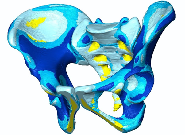
A series of 150 pelvic CT scans (50 European females and 50 males, 20 Asian females and 30 males) were post-processed with gender and ethnicity specific 3D statistical models of the pelvic ring generated.
A workflow for symmetry analysis has been elaborated for identification of symmetry patterns in given analysed CT cases. The pelvic ring analysis resulted in a symmetric complex configuration with regard to the bony surface and its internal configuration. Distinct anatomical sites were identified where asymmetry patterns were typically located.
-
PublicationHandrich K, Kamer L, Mayo K, Sawaguchi T, Noser H, Arand C, Wagner D, Rommens PM. Asymmetry of the pelvic ring evaluated by CT-based 3D statistical modeling. J Anat. 2020;epub Dec 31. https://doi.org/10.1111/joa.13379
-
Partner
Rommens PM (Prof), University Medical Center, Mainz, Germany
Mayo K (Prof), University of Washington School of Medicine, Seattle, USA
Sawaguchi T (Prof), Toyama Municipal Hospital, Toyama, Japan
Arand C (MD), University Medical Center, Mainz, Germany
Wagner D (MD), University Medical Center, Mainz, Germany