Histology, tissue morphology and microscopy
Highly-skilled tissue processing, imaging, and analysis
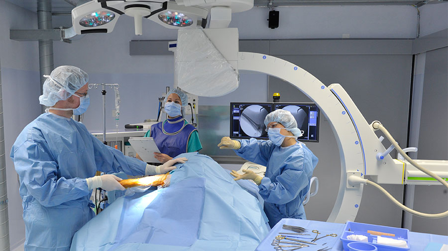
Study design
- Assistance in project planning and experimental set-up based on scientific question
- Evaluation of samples, photodocumentation, and report writing
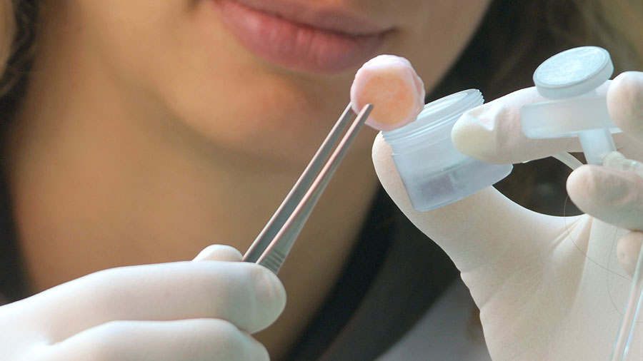
Histological preparation and techniques
- Undecalcified bone histology with or without implants (eg, metal, calcium phosphates, polymers)
- Sectioning of resin embedded samples with specialty equipment
- Contact radiography
- Decalcification of tissues
- Paraffin embedding and sectioning
- Cryosectioning
- Immunohistochemistry (with protocol planning)
- Routine to special stains (for resin and non-resin embedded samples)
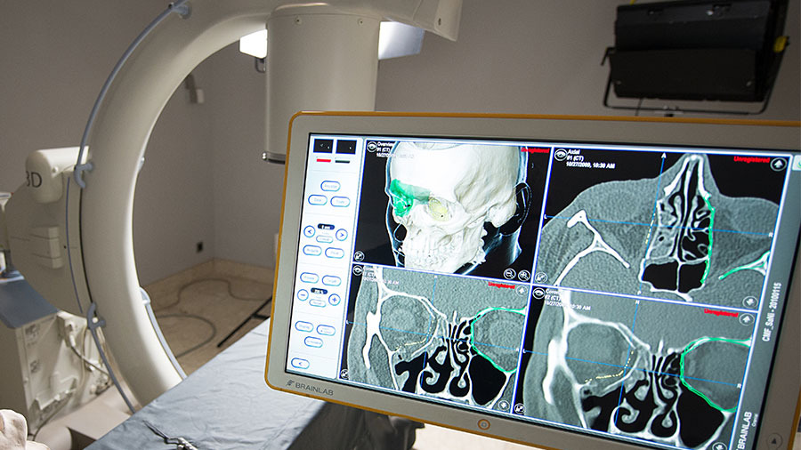
Microscopy and image analysis
- Brightfield light microscopy for sections with or without implants
- Epifluorescent microscopy for fluorochrome labelled sections (eg, bone labels, marked antibodies)
- Macrophotography for overview images of larger samples
- Confocal laser microscopy for optical sectioning
- Scanning electron microscope equipped with Energy Dispersive X-Ray Spectroscopy (EDX or EDS)
- White light profilometry for surface characterization (eg, roughness values)
- Quantitative analysis (eg, lengths, areas, numbers) on histological samples
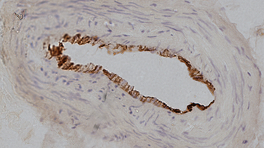
CD31 immunohistochemical stain showing a blood vessel, of a decalcified, cryosectioned coracoclavicular ligament
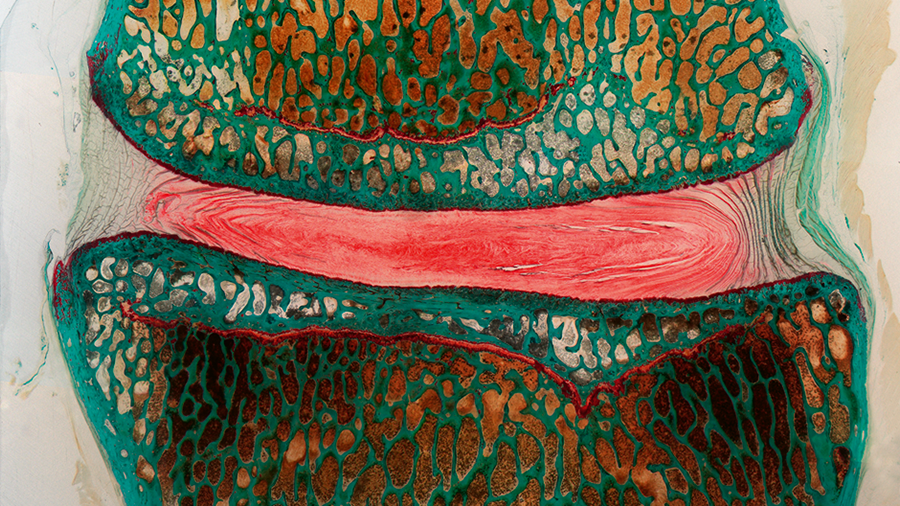
Safranin O/Fast Green staining: resin embedded sheep lumbar disc
Qualifications
- Certified vet. pathologist
Certification
- ISO 9001
- Good Laboratory Practice (GLP)
Contact
Ulrich Bentz
AO Research Institute Davos
Clavadelerstrasse 8
7270 Davos Platz
Switzerland
Tel.: +41 81 414 23 26