
Creation of a statistical form model of the cortical femur with tools for automatic feature extraction and fracturing (cortFemSFM)
Background
Bone models are required in many projects on preclinical research and development, however, the traditional approach for their acquisition is expensive and time consuming.
In some cases, computerized bone models can be used as alternative approach. For example, the analysis of individual anatomy or variation patterns of a given population sample can facilitate surgical decision making, preoperative planning and development of implants for orthopedic trauma care.
Goal
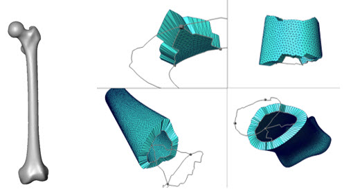
To develop a workflow and generate a 3D statistical form model of the femur and its cortex, containing inherent anatomical information about the femoral size, cortical thickness, shape and length.
Results
New modules under development include 3D modeling of the femoral cortex and fracture pattern, including fracture segments and a module for morphometric analysis (eg., for length and angle measurements). Currently, 40 CT scans of the femur have been segmented, and a workflow has been developed to produce a 3D statistical form model of the femur and its cortex. The procedure includes a fracture tool that requires outlining the fracture lines to generate fracture segments thereof.