
Cone-beam computed tomography – a fast and promising technique for microstructural imaging in clinical practice
Background
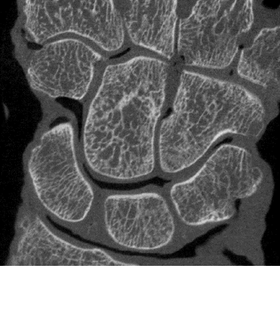
Obtaining high-resolution CT scans for clinical applications is challenging, However, it could help better understand and treat such bone diseases as osteoporosis. High-resolution peripheral computed tomography (HR-pQCT) is considered the best technique in vivo. However, a breakthrough for clinical practice is lacking due to a relatively long acquisition time, which inhibits scanning of large field of view (FOV) in vivo. A promising alternative is the high-resolution cone-beam computed tomography (CBCT), which is already the gold standard in many dental and maxillofacial applications. The top high-resolution CBCT scanners on the market (eg., Newtom 5G) feature a fast scanning time (18 à 31s), a large FOV (12x12x8cm3) and a low radiation dosage, in addition to a high resolution (voxel size down to 75µm). Yet, CBCT is impaired by the presence of image artefacts that reduce image contrast, leading to it being currently used for qualitative evaluation only.
Goal
To determine whether CBCT can be enhanced by means of artefact correction algorithms and advanced segmentation techniques in order to be used to visualize and quantify both bone microstructure and mechanical parameters in clinical practice.
Results
CBCT enables fast scanning of large FOV of extremities at high spatial resolution. In addition, the enhanced CBCT images had a very comparable accuracy with HR-pQCT when quantifying bone microstructural and mechanical parameters. When enhanced, high-resolution CBCT is an attractive and promising imaging tool that can be used in the clinical treatment of several bone and joint diseases.
Fund:
KU Leuven internal funding and the Swiss National Supercomputing Centre under project ID 593, ARI funding: EUR 7'000, Period: 2019
-
Publication
Mys K, Varga P, Gueorguiev B, Hemmatian H, Stockmans F, van Lenthe GH. Correlation between cone-beam computed tomography and high-resolution peripheral computed tomography for assessment of wrist bone microstructure, J Bone Miner Res. 2019,34:867-874
Mys K, Varga P, Gueorguiev B, Hemmatian H, Stockmans F, van Lenthe GH. Reply Letter to the editor: Clinical in Vivo Assessment of Bone Microarchitecture with CT Scanners: An enduring challenge, J Bone Miner Res. 2019
Mys K. Cone-beam computed tomography is a fast and promising technique for microstructural imaging in clinical practice, PhD thesis, 2019
-
Presentation
Mys K, Zhang G, Stockmans F, Wyers CE, van den Bergh JPW, van Lenthe GH. X-Ray Scattering Is Limited in Cone-Beam Computed Tomography of Extremities. 2019 QMSKI, Lake Louise, Canada (oral)
Mys K, Varga P, Stockmans F, Gueorguiev B, Wyers CE, van den Bergh JPW, van Lenthe GH. Cone-Beam Computed Tomography as a Fast Alternative for High-Resolution Peripheral Computed Tomography. 2019 QMSKI, Lake Louise, Canada (oral)
Mys K, Varga P, Stockmans F, Gueorguiev B, Wyers CE, van den Bergh JPW, van Lenthe GH. Influence of the Analysis Software to Calculate Direct Bone Structural Parameters. 2019 QMSKI, Lake Louise, Canada (poster)
Mys K, Varga P, Stockmans, F, Gueorguiev B, Neumann V, Vanovermeire O, Wyers CE, van den Bergh JPW, van Lenthe, GH. Cone-beam CT as a fast and promising technique to assess the microstructure of distal radii in clinical practice. 2019, ESB, Vienna, Austria (oral)
-
Partner
Van Lenthe GH (Prof), KU Leuven, Leuven, Belgium
Stockmans F (Prof), KU Leuven Kortrijk, Kortrijk, Belgium