Can arthroscopic techniques contribute to further development of PLC injury treatment?

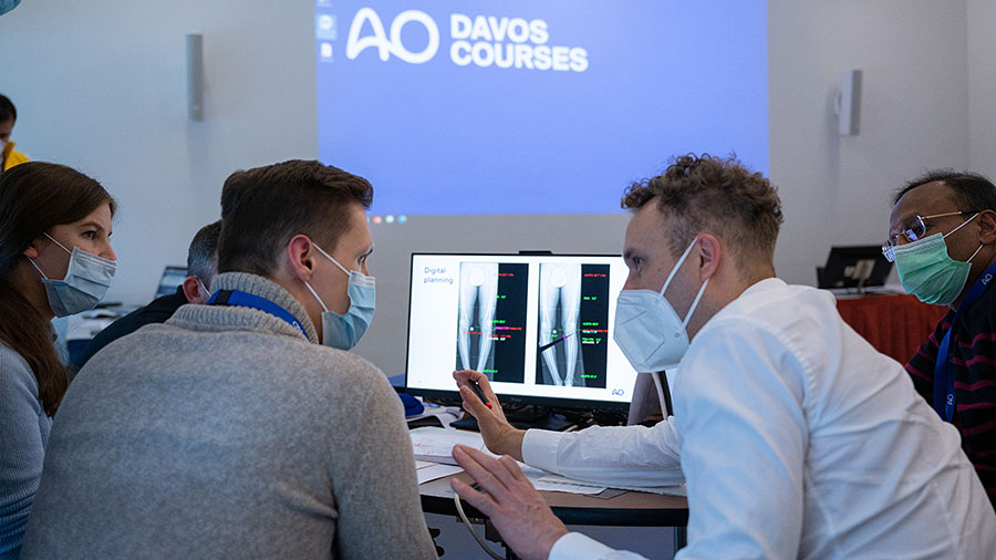
Dr Matthias Krause (right) at the first AO Sports Advanced Course--Knee Injuries and Deformities, AO Davos Courses, 2021
By: Matthias Krause, MD, PhD
Senior physician, specialist in orthopedics and trauma surgery
Department of Trauma and Orthopaedic Surgery, University Medical Center Hamburg-Eppendorf (UKE), Hamburg, Germany
Injuries to the posterolateral corner (PLC) are common in posterior instabilities of the knee but may also be found in anterior cruciate ligament (ACL) injuries. The PLC consists of the popliteus muscle/tendon unit (PLT), the popliteofibular ligament (PFL), the fabellofibular ligament, and the popliteomeniscal fibers (arcuate complex [AC]).
PLC injury may lead to increased posterior, external rotational, and/or varus instability and, thus, provide the biomechanical basis for graft failure following cruciate ligament reconstruction due to missing load sharing support. Accordingly, anatomical reconstruction techniques have been proposed to stabilize the PLC better than nonanatomical techniques.
Arthroscopic procedures for the treatment of posterolateral instabilities
Recently, arthroscopic procedures have gained popularity for the treatment of posterolateral instabilities. Some targeted techniques, such as the arthroscopic popliteus bypass, are designed to address isolated rotational instabilities (eg, Fanelli type A). Other technical descriptions have focused on arthroscopic techniques for complex anatomical PLC reconstruction. Although these techniques show promising biomechanical and clinical results, they are technically demanding and require a fundamental understanding of the arthroscopic posterolateral anatomy.
Fibular tunnel placement
The accurate and reliable anatomical tunnel placements for arthroscopic PLC reconstruction at the femoral and tibial sides have already been described. With respect to the fibular tunnel, “anatomical” typically refers to the single-tunnel fixation from anterolateral inferior to posteromedial superior, mimicking the fibular collateral ligament (FCL) and PFL footprints in contrast to a nonanatomical anteroposterior tunnel trajectory. As the PFL is the most important static stabilizer against tibial external rotation, exact and reliable fibular tunnel placement is crucial, especially as the fragile fibular head is prone to fracture, graft loosening, and subsequent laxity. With the recent progress of arthroscopic PLC reconstruction techniques, anatomical fibular tunnel placement might be improved but has not yet been validated, especially in direct comparison to open reconstruction. In a recent study, the accuracy of arthroscopic fibular tunnel placement was compared with the open technique in terms of their safe distance to surrounding cortical edges of the fibular head.
In that study’s retrospective analysis, reasonable soft-tissue and bony landmarks—which can be identified by either arthroscopy, fluoroscopy, or open surgery in anatomical fibular tunnel placement—were identified. Subsequently, the authors compared the ideal fibular tunnel created with arthroscopy and in a standard open technique from anterolateral inferior to posteromedial superior with a 2 mm K-wire in a cadaver study.
Optimal fibular tunnel entry and exit
Based on magnetic resonance imaging (MRI) measurements, the optimal fibular tunnel entry on average should be 14.50 (±2.18) mm distal to the tip of the fibular head and 10.76 (±1.37) mm posterior to the anterior edge of the fibula. With respect to the arthroscopic visualization of the PLC, the ideal fibular tunnel exit point was located 2.06 (±2.42) mm above the superior border of the soleus muscle and 12.89 (±2.35) mm below the tip of the fibular head (fibular styloid). The peroneal nerve was located 19.96 (±4.94) mm distally to the ideal tunnel exit point. Angulation of the fibular tunnel was 23.15 (±4.14) degrees rotated internally with respect to the tangent line of the dorsal tibial condyles (axial plane) and 23.40 (±3.30) degrees pivoted upward with respect to the sagittal/coronal plane. Most importantly, arthroscopic anatomical tunnel application was more reliable in creating a safe distance of the tunnel center to the closest cortical edge than open tunnel placement, which showed a tunnel malposition in five out of eight cases based on keeping an 8 mm distance. As the PFL originates right at the tip of the fibular head, the fibular tunnel exit should not be placed at the anatomical PFL footprint but more distally to prevent fibular head fracture.
Conclusion
The authors concluded that arthroscopic techniques can make an important contribution to the further development of the treatment of PLC injuries. The two most important criticisms of arthroscopic PLC reconstruction include nonanatomical tunnel placement and that neurovascular injuries cannot be supported by anatomical findings of the present study.


