
Research grants
Global and regional funding and training opportunities for Trauma research excellence
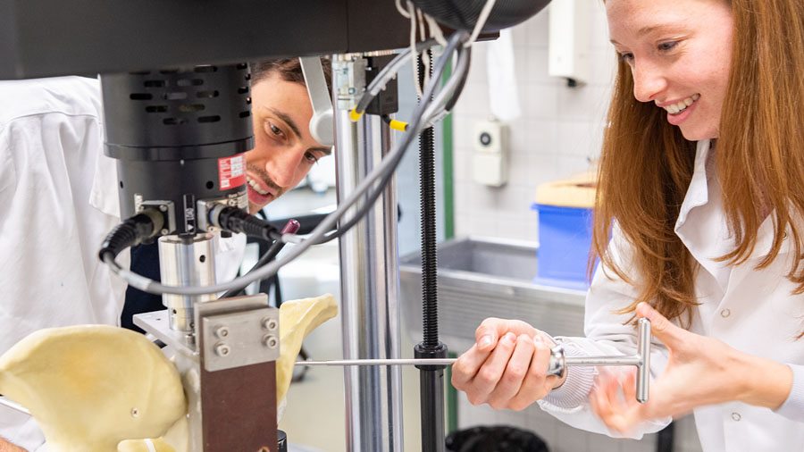
As part of its commitment to supporting individual research career development for clinicians, AO Trauma offers research grants that fund clinical, basic, and applied research focused on solving highly relevant problems in trauma and musculoskeletal care.
These opportunities are available to AO Trauma clinicians from all global regions—Europe and Southern Africa (ESA), Middle East and Northern Africa (MENA), Asia Pacific (APAC), North America (NA), and Latin America (LATAM).
In addition to traditional research grants, AO Trauma is advancing structured training opportunities for young investigators, including the development of training grants, enhanced research mentorship, and innovative surgical training courses that promote best practices in preclinical and clinical research. These initiatives aim to foster a culture of excellence, innovation, and evidence-based care across the trauma community.
Funding opportunities
Have a bold idea in trauma research?
Apply for funding through AO Trauma’s open grant calls and contribute to advancing evidence-based care in musculoskeletal trauma.
-
AOTrauma_S_APAC_2025-01673: Development of innovative implant for Hoffa fracture
Principal Investigator: Harjot Gurduatta, Shri Akal Purakh Hospital, India
Clinical research
Abstract:
Abstract Hoffa fractures, once considered simple posterior condylar injuries, have shown complex comminution patterns and multiple fracture lines, necessitating improved fixation methods.
This proposal outlines the development of a novel, anatomically contoured implant system specifically designed for Hoffa fractures. The system includes a curvilinear base plate and a multi-pronged buttress component to stabilize articular and metaphyseal fragments.
Biomechanical testing will be conducted on dry bones, cadavers, and patient models, building on an initial successful pilot in five lateral Hoffa cases.
The implant aims to enhance fracture stability, enable early mobilization, and offer superior tissue coverage due to its low-profile, moldable design. The one-year project includes design, prototyping, clinical testing, and biomechanical validation, with an estimated budget of USD 15,000.
-
AOTrauma_S_APAC_2025-01707: Assessing pelvic stability during physiological loading in cadaveric models
Principal Investigator: Qinxiang Shant Sin, Ministry of Health Holdings, Singapore
Translational research
Abstract:
Pelvic ring injuries are caused by severe trauma such as road traffic accidents and crush injuries. These patients may be in a foreign country and may wish to be treated back in their home country. Repatriation back is a challenge as pelvic ring injuries are severe and patients may not be able to tolerate sitting upright or lying in a reclining seat in a long flight. Lying flat may require additional logistics and costs which may be unfeasible for the patient.
Based on current literature, there is a lack of established protocols that support the safety of such patients to sit or stand. This leaves an uncertainty in patient's positioning if repatriation was required. The decision is left to the discretion of the treating physician, of which some may disallow repatriation due to perceived pelvic instability. This study involves 6 cadaveric pelvis models that tests the tension and durability of remaining ligaments when one or more ligaments have been damaged. Tension meters will be placed on all major pelvic ligaments (pubic symphysis, sacrotuberous, sacrospinous, anterior sacroiliac joint, posterior sacroiliac joint ligaments). The pelvic specimen will be clamped and fixed in sitting and standing positions. A physiological load will be applied to the models and the baseline tension force in these tension meters will be measured. Subsequently, each ligament will be surgically sectioned sequentially from anterior to posterior to mimic ligament rupture. The maximum physiological load to keep the pelvis stable during each position will be recorded.
This aims to assess the pelvic stability in a physiological standing or sitting position. This study can be extrapolated into a clinical setting to guide physicians in deciding the positioning of patients for repatriation or medical transfers. The results obtained can also be used to craft rehabilitation protocols for patients with conservatively treated pelvic ring injuries to start mobilization (sitting or standing) earlier.
-
AOTrauma_S_APAC_2025-01845: Anthropometric Measurement for Estimated Femoral Nail Length
Principal Investigator: Mohd Afiq Bin Muhamed Fuad, Faculty of Medicine and Health Sciences, Universiti Putra Malaysia, Malaysia
Clinical research
Abstract:
Choosing the correct intramedullary nail length is critical for the successful fixation of femoral-shaft fractures. Current standards rely on X-ray measurements or the uninjured femur?methods that are impossible in bilateral fractures, expose patients to radiation, and are difficult in obesity or pregnancy. We propose a simple anthropometric alternative: using palpable fibula length to estimate maximum femoral nail length (EFNL).
Aims: Quantify the correlation between clinical fibula length and EFNL. Derive a predictive formula for EFNL based solely on fibula length. Verify measurement reliability between observers. Explore whether age, sex, height or weight influences the correlation. A reliable, tape-measure method eliminates unnecessary imaging, shortens operating-room preparation, lowers costs, and offers a safe option for polytrauma and radiation-sensitive patients.
Materials & Methods Design: single-centre, cross-sectional study.
Setting: Orthopaedic clinic and ward, Hospital Sultan Abdul Aziz Shah (HSAAS).
Population (P): 130 ambulatory patients, 12–60 y, without lower-limb deformity.
Intervention (I): Clinical measurement of fibula length (head styloid – lateral malleolus) by two blinded observers.
Comparison (C): Clinically estimated EFNL (greater trochanter – line perpendicular to superior patella).
Outcome (O): Strength of correlation, predictive equation, inter- and intra-observer reliability.
Time frame (T): Single measurement session per patient (Jun 2025–July 2026).
Analysis: Spearman correlation, zero-intercept linear regression, multiple regression for covariates, and intraclass correlation coefficient (ICC) for reliability.
Expected Results: Fibula Length have significant correlation with EFNL.
Long-term Goal: To integrate fibula-based EFNL estimation into routine orthopaedic practice especially those with bilateral femoral injuries or conditions that preclude conventional measurement.
-
AOTrauma_S_APAC_2025-01852: Clinical efficacy of 3D Ti- and Mg-implants for repairing peri-articular defects
Principal Investigator: Bingchuan Liu, Peking University Third Hospital, China
Clinical research
Abstract:
The treatment of bone defects in the peripheral joints of the limbs is a challenging problem in orthopaedic clinical practice, and traditional methods are unable to achieve efficient and safe repair of complex bone defects. The use of emerging 3D printing technology for the treatment of bone defects offers the advantages of personalisation and precision.
Our research team has successfully treated over 100 patients with limb bone defects using 3D-printed titanium alloy bone implants, accumulating extensive experience in the process. Additionally, we have completed in vitro and in vivo performance tests for 3D-printed magnesium alloy bone implants and obtained the necessary ethical approval for clinical trials, marking a pioneering exploration not yet reported in domestic or international literature.
Based on this foundation, this project will fully leverage the advantages of prior research and practical experience to conduct an in-depth comparison of the clinical efficacy differences between using 3D-printed titanium alloy and magnesium alloy bone implants for repairing limb joint bone defects.
The focus will be on customised design and production of bone implants, stable fixation patterns, characteristics of new bone regeneration, degradation patterns of magnesium alloy implants, joint function rehabilitation, and complications.
The research findings will help comprehensively and deeply address the challenges encountered in the clinical application of 3D printing technology in orthopaedics, provide new materials and technologies for the clinical treatment of bone defects around the joints of the limbs, and lay the foundation for the clinical translation of products and the initiation of multi-center studies.
-
AOTrauma_S_APAC_2025-01865: AI-Driven 3D Reconstruction of Pediatric Supracondylar and Lateral Condyle Humerus
Principal Investigator: Shigeki Ishibashi, Hiroshima University Hospital, Japan
Clinical research
Abstract:
Background: Assessing complex pediatric elbow fractures, particularly those involving the epiphyseal plate, is challenging with 2D radiographs alone. While computed tomography (CT) provides essential 3D detail, it poses risks of radiation exposure and higher costs in pediatric care. Deep learning can reconstruct 3D models from X-rays, but a clinically validated model for the unique anatomy of the pediatric elbow is a critical unmet need.
Objective: This study aims to develop and validate a deep learning algorithm that accurately reconstructs 3D models of pediatric supracondylar and lateral condyle humerus fractures from biplanar radiographs, with the goal of creating a reliable alternative to pre-operative CT scans.
Methods: Using a retrospective, multicenter dataset of 250 patients with paired radiographs and CT scans, we will train a deep learning model featuring a dual-stream 2D encoder and a 3D U-Net decoder to generate 3D bone geometry. Model accuracy will be rigorously validated against CT-derived ground-truth models using metrics such as the Dice Similarity Coefficient (DSC) and mean surface distance.
Anticipated Results: We hypothesize that the algorithm will reconstruct the fracture geometry with a mean surface error of less than 2.0 mm and achieve a DSC greater than 0.85. The resulting 3D models are expected to provide clinically reliable anatomical information for accurate diagnosis and surgical planning.
Conclusion & Significance: A successful model would offer a novel diagnostic tool to improve surgical planning for complex pediatric elbow fractures while significantly reducing the need for CT scans and their associated radiation and costs. Furthermore, this technology could enable 3D assessment of fracture healing from routine follow-up radiographs, establishing a new paradigm in pediatric orthopedic imaging.
-
AOTrauma_S_APAC_2025-01885: AI-CT Quantification of Muscle Recovery in High-Energy Fracture Patients
Principal Investigator: Suguru Yokoo, Fukuyama City Hospital, Japan
Clinical research
Abstract:
Background: High-energy pelvic and femoral fractures precipitate rapid lower-limb muscle atrophy and fatty infiltration, delaying rehabilitation and prolonging disability. Current CT-based workflows rely on manual slice measurements that are subjective, slow, and poorly reproducible, and no automated volumetric tool exists to guide early, muscle-specific therapy.
Aim: This study will (1) apply a Bayesian 3D U-Net pipeline to quantify immediate postoperative (< 7 days) volume loss in key lower-limb muscles of 150 adult fracture patients, and (2) predict and internally validate their recovery trajectories at 3 and 6 months.
Relevance: Early identification of muscle groups at risk for delayed regeneration enables personalized rehabilitation, improves functional gains, and reduces inpatient stays.
Methods: In this retrospective cohort (2018–2025), we will automatically segment quadriceps, hamstrings, abductors/adductors, and flexors on serial CTs (preoperative, postoperative ≤ 7 days, 3 month, 6 month). We will quantify absolute and relative volume changes, stratify by fracture type, age, and sex, and correlate muscle‐volume changes with change in Barthel Index at 3 months, length of stay (LOS), and return‐to‐home rate.
Expected Results: We anticipate identifying at least two muscle groups with early volume loss ≥ 15 %, and that such early losses will independently predict a ≥ 10-point lower Barthel Index at 3 months and a 20 % longer LOS compared with patients without significant early loss.
Long-term Goal: To integrate CT-derived recovery maps into standard post-fracture care protocols, thereby enabling data-driven, individualized physiotherapy regimens.
PICOT:
P—Adults with high-energy pelvic/femoral fractures;
I—AI-CT segmentation plus targeted early rehab;
C—Historical usual care;
O—Muscle‐volume change vs. Barthel Index change at 3 months (primary); secondary: LOS and return‐to‐home rate;
T—Baseline through 6 months. -
AOTrauma_S_APAC_2025-01889: Cutting Risk Before the Cut: Fructosamine’s Role in Infection Prediction
Principal Investigator: Ashok Ramanujam, Mahatma Gandhi Medical College & Research Institute, India
Clinical research
Abstract:
Introduction and Background: Postoperative infections remain a significant complication in orthopaedic trauma surgery, particularly among patients with impaired glucose metabolism. Glycemic control is a known determinant of surgical outcomes, yet traditional markers such as HbA1c reflect long-term glucose control and may not capture rapid changes prior to surgery. Serum fructosamine, a marker of short-term glycemic status (2–3 weeks), has shown emerging potential as a predictive biomarker for postoperative infections but is underutilized and under-researched in the orthopaedic trauma setting. This study aims to fill this critical knowledge gap.
Aim: The primary aim is to evaluate the association between preoperative serum fructosamine levels and the incidence of postoperative infections in orthopaedic trauma patients.
Objectives:
- Measuring serum fructosamine levels preoperatively in patients undergoing trauma surgery.
- Monitoring and recording postoperative infections over a defined follow-up period.
Materials & Methods:
Design: Prospective, observational cohort study.
Study Period: September 1, 2025–September 30, 2027.
Population (P): Adult patients (≥18 years) undergoing orthopaedic trauma surgery.
Intervention (I): Measurement of serum fructosamine levels within 48 hours prior to surgery.
Comparison (C): Outcomes compared across patients with normal vs. elevated fructosamine; secondary comparison with HbA1c levels.
Outcome (O): Incidence of surgical site infections (superficial and deep), including periprosthetic joint infections where applicable.
Time (T): Follow-up period of 90 days postoperatively for infectionExpected Results: We anticipate that elevated serum fructosamine levels will be significantly associated with higher rates of postoperative infections.
-
AOTrauma_S_APAC_2025-01897: Evaluating Humeral Stem Length on Implant Mechanics in Total Elbow Arthroplasty
Principal Investigator: Merrill Lee, Singapore General Hospital, Singapore
Translational research
Abstract:
Expanding indications subjects the Total elbow arthroplasty (TEA) to increased mechanical stresses leading to potential complications such as loosening, implant component fracture and peri-prosthetic fracture. This computational analysis aims to investigate the overall biomechanical performance of the total elbow implant in response to varying implant lengths. By utilising different implant lengths, we hope to demonstrate its effect on the bone and implant stresses and identify changes in the load transfer pattern. We hypothesize that modifying the implant length will result in changes in the bone and implant stresses. We also hypothesize that by using the optimum implant length, we can restore the bone stresses in the bone-prosthesis assembly to its intact state.
Three synthetic humerus and ulna sawbones as well as its corresponding digital models to perform computational analyses will be used. Distal total elbow implants of varying lengths will undergo laser scanning to obtain the three-dimensional (3D) digital models which will then be used to generate bone-prosthesis assemblies. The models will be exported to a finite element solver software for simulation. Three load cases at specified flexion angles with the corresponding axial loads calculated with respect to joint reaction force and flexion angle as well as a torsional moment will be applied for both experimental and computational analysis. Strain readings from experimental testing and computational analyses will be compared at respective locations to validate the intact model. The stresses and strains across the bone-implant assemblies with varying implant lengths are evaluated after separating the cortical bone, cancellous bone and implant.
The following study will possibly guide clinicians in determining the optimum implant length with improved biomechanical performance for their patients and potentially reduce the incidence of mechanical complications.
-
AOTrauma_S_APAC_2025-01908: Ultrasonic automated screw tunnel depth gauge for orthopaedic surgery
Principal Investigator: Kia Teng Lim, National University Hospital, Singapore
Translational research
Abstract:
Our research aims to develop a novel depth gauge that integrates guiding sleeves, an embedded ultrasound probe, and an electronic screw length measurement system to enhance accuracy in orthopaedic screw placement, particularly in trauma surgery.
Traumatic injuries remain a major global health burden, with fracture fixation being one of the most common surgical interventions. Screws are widely used not only in trauma care but also in procedures like total hip replacements and limb deformity corrections.
Current screw placement relies on manual depth gauges and memory of the drilled trajectory after removal. Inaccuracy in screw placement and screw length measurements using depth gauges remains an obstacle—studies reveal that ideal screw placement is achieved in only 49.2% of cases, with no difference between senior and junior surgeons. Incorrect screw lengths compromise fixation integrity—short screws may fail to engage the far cortex, while long screws risk damaging soft tissues or neurovascular structures. Repeated screw removals due to miscalculations can prolong surgeries, increase costs, and damage bone integrity. Repeated intra-operative imaging to confirm placement accuracy exposes both patients and surgical teams to ionizing radiation, which poses health risks and extends operative time.
Our multidisciplinary team comprising orthopaedic surgeons, radiologists, and biomedical engineers will collaborate to design and prototype this new device. By eliminating the need for the traditional hook mechanism, the ultrasound-enhanced depth gauge will allow accurate screw length determination with real-time feedback. We will evaluate the device's performance against standard tools using both sawbone and cadaveric models across various surgical experience levels, aiming to establish a safer, more efficient, and cost-effective standard for orthopaedic screw placement.
-
AOTrauma_S_APAC_2025-01918: Optimal screw trajectory for antegrade infraglenoid screw insertion in scapula
Principal Investigator: Whee Sung Son, Orthopaedic Department, Yeungnam University, Republic of Korea
Clinical research
Abstract:
Background: Scapular fractures involving the glenoid articular surface critically affect shoulder function and long-term outcomes. Despite this importance, there is no established guidance on optimal screw trajectory for fixing the inferior glenoid fragment. This project addresses that gap by defining the "antegrade infraglenoid screw" trajectory—a concept inspired by the infraacetabular screw in pelvic surgery to achieve long, stable fixation and improve surgical precision.
Aims: To determine the optimal screw trajectory and plate position for maximizing purchase in the coracoid using CT-based 3D simulation. Objectives will be achieved by modeling screw corridors and plate positions on detailed scapular 3D reconstructions.
Relevance: The study will provide actionable guidance for surgical planning in glenoid fractures, improving fixation strength, reducing complications, and leading to better functional outcomes.
Materials and Methods: Shoulder CT scans (complete scapula, no fractures/deformities) will be reconstructed into 3D models. Virtual screw corridors will identify optimal starting points and angles from inferior glenoid to coracoid. Plate fixation simulations at 6, 6:30, and 7 o'clock positions on the glenoid rim will evaluate cross-sectional purchase areas for each screw hole within typical angulation ranges.
Preliminary Results: Analysis of 10 scapular CT scans showed the optimal antegrade infraglenoid screw trajectory averaged 9.16 ± 3.46° medially (coronal) and 28.63 ± 3.46° anteriorly (sagittal), with a mean corridor area of 159.37 ± 61.56 mm². The first screw hole at the 6 o’clock plate position provided the greatest purchase volume in the coracoid (mean 928.48 ± 213.68 mm³), outperforming other positions and holes, suggesting it is the most favorable for reliable fixation.
Long-Term Goal: To establish evidence-based surgical techniques for scapular fractures that improve fixation reliability, reduce failure rates, and enhance patient outcomes.
-
AOTrauma_S_APAC_2025-01921: Why Does VALPP Fail? A Multi-dimensional Analysis of Comminuted Patellar Fracture
Principal Investigator: Jeonghyun Koh, Ajou University Hospital, Republic of Korea
Clinical research
Abstract:
Problem and Background: The Variable Angle Locking Patellar Plate (VALPP) has emerged as a promising solution for complex patellar fractures, yet recent evidence reveals concerning 40% mechanical failure rates in AO/OTA 34-C3 fractures with inferior pole comminution. This represents a critical clinical problem as inferior pole involvement creates concentrated tensile forces that challenge current fixation strategies.
Significance and Innovation: This study addresses a significant knowledge gap by implementing a novel multi-dimensional analysis framework combining statistical risk profiling, 3D morphological failure mapping, and comparative performance analysis. The innovative approach will identify specific fracture morphologies predicting failure and visualize failure patterns through advanced 3D heatmap technology.
Aims and Methods: We will conduct an ambispective comparative cohort study analyzing 100 patients with AO/OTA 34-C3 patellar fractures, comparing standalone VALPP versus competing plate with mini-plate augmentation. The study employs three analytical dimensions: multivariate statistical analysis for risk factor identification, 3D CT-based fracture mapping for failure pattern visualization, and head-to-head comparative analysis for treatment optimization.
Expected Results: We hypothesize that augmented fixation strategies will reduce mechanical failure rates from 40% to ?15% in inferior pole comminuted fractures, with 3D analysis revealing characteristic failure signatures at the inferolateral patellar aspect.
Clinical Impact: This research will provide surgeons with evidence-based risk stratification tools, visual failure pattern atlases, and optimized treatment algorithms, potentially preventing numerous revision surgeries across the Asia-Pacific region.
-
AOTrauma_S_APAC_2025-01934: LC2 screw increase the fixation stability over the plate osteosynthesis FFP IIIA
Principal Investigator: Sangroc Han, Kyungpook National University Hospital, Republic of Korea
Clinical research
Abstract:
Fragility fractures of the pelvis (FFP) pose a significant clinical challenge in the elderly population due to osteoporosis-induced structural weaknesses. Among FFP, type IIIa fractures affecting the posterior pelvic ring necessitate robust fixation strategies to prevent fixation failure in osteoporotic bone. Standard plate osteosynthesis offers reliable fixation but is limited by implant loosening and screw withdrawal risks in poor-quality bone. Recent evidence suggests that a combined fixation using a lateral compression (LC2) screw along with an anterior plate could offer superior biomechanical stability. The primary aim of this study is to compare the biomechanical effectiveness of a novel combined anterior plate and LC2 screw fixation (nail-plate construct, NPC) versus standard plate-only fixation (PO) in osteoporotic FFP type IIIa fractures.
The project will utilize synthetic osteoporotic hemipelvis models subjected to standardized fracture creation. Each specimen will be tested biomechanically using cyclic axial loading protocols at AO Research Institute, Davos. Outcomes including stiffness, displacement under load, angular movement, and failure modes will be assessed. This study expects to demonstrate superior stability of the NPC fixation, potentially reducing complications from fixation failure.
Long-term, these results aim to establish a foundation for improved surgical approaches in geriatric pelvic fracture management.
PICOT Summary
P : Elderly patients with fragility fractures of the pelvis (FFP) type IIIa
I : Combined fixation using anterior plate and LC2 screw
C : Plate-only fixation
O : Biomechanical stability, reduced implant failure
T : Single-year in vitro study (July 2025–June 2026) -
AOTrauma_S_APAC_2025-01946: Biomechanical Analysis of Fragility Fractures of the Pelvis
Principal Investigator: Masahiro Kawagishi, Oita University Hospital, Faculty of Medicine, Japan
Translational research
Abstract:
Fragility fractures of the pelvis (FFPs) are increasingly observed in the aging population and are strongly associated with osteoporosis. Despite growing clinical awareness, the biomechanical mechanisms underlying FFPs and the optimal fixation strategies remain poorly understood.
This study aims to identify fixation methods that are both biomechanically sufficient and minimally invasive, tailored to each FFP classification type. We will construct patient-specific finite element (FE) models of osteoporotic pelvic bones using CT data and simulate typical FFP patterns by introducing linear fracture lines according to the Rommens classification. Various fixation techniques—including trans-sacral screws, LC2 screws, and triangular fixation methods such as TIRF and SPF—will be evaluated for mechanical stability through stress distribution, failure element analysis, and displacement at the fracture site. For type IV FFPs in particular, the study will examine which fixation provides the most stable yet least invasive construct.
To validate the simulation findings, we will replicate selected FE models using physical sawbone experiments and measure interfragmentary motion and load to failure. These results will be compared to FE outcomes to enhance clinical applicability. By determining fixation strategies that are necessary and sufficient for each FFP subtype, this study aims to reduce postoperative instability and reoperation risk, and contribute to the development of evidence-based surgical treatment protocols.
-
AOTrauma_S_APAC_2025-01949: A Score Driven Framework to Guide Decisions in Geriatric IT Fractures
Principal Investigator: Nalli Uvaraj Sanjeev Kumar, Sri Ramachandra Institute of Higher Education and Research, India
Clinical research
Abstract:
Intertrochanteric hip fractures in the elderly present a unique surgical dilemma, balancing the goals of early mobilization against the challenges of poor bone quality and complex fracture morphology. While internal fixation remains the standard treatment for most cases, primary hemiarthroplasty is increasingly considered in selected unstable, osteoporotic fractures to avoid mechanical failure and prolonged immobilization. However, the decision to choose arthroplasty over fixation has traditionally relied on subjective clinical judgment, leading to variability in practice and complications.
To address this gap, there is a growing interest in developing objective, score-based frameworks that systematically integrate patient factors, fracture stability, and bone quality.
Such tools aim to guide surgeons toward evidence-based, individualized treatment choices, improving functional outcomes and reducing complications. We intend to explore the development and application of an objective, score-based guide to assist in selecting hemiarthroplasty for intertrochanteric hip fractures.
While internal fixation remains the standard for most intertrochanteric fractures, primary hemiarthroplasty is selectively used in certain high-risk or unsuitable cases.
-
AOTrauma_S_APAC_2025-01954: Comparison of biomechanics fixation for sacroiliac joint disruption
Principal Investigator: Nachapan Pengrung, Ramathibodi Hospital, Thailand
Clinical research
Abstract:
This leads to the research question of whether using the dual coaxial combi-holes anterior plate with four cortex screw catching instead of traditional fasion can enhance the strength of sacroiliac joint fixation compared to sacroiliac screws.
-
AOTrauma_S_LATAM_2025-1-01365: Fijación mínima invasiva con placa en fracturas
Principal Investigator: Daniel Navarro, Vergara Paraguay, Paraguay
Clinical research
Abstract:
Introduccion: La diferencia entre de las lesiones en la edad pediátrica con las de los adultos son numerosas, y requieren manejos distintos, ya que las características del hueso, el tiempo de consolidación y el contexto emocional son diferentes. En la actualidad, las indicaciones de cirugía en los pacientes pediátricos han ganado terreno, sobre todo en las fracturas femorales en los niños mayores de 4 años, manteniéndose la indicación de tratamiento conservador en las fracturas de tibia, con pocas indicaciones absolutas de cirugía en las lesiones a este nivel. Numerosas son las ventajas que se citan defendiendo las indicaciones de cirugías en la población infantil. Es así, como el uso de clavos endomedulares (ESIN: Elastric Stable Intramedulary Nail) en las fracturas de los huesos largos de la extremidad inferior se ha mostrado como patrón de oro, pero su uso común, acarreo la aparición de complicaciones. En este sentido, es que el uso de la Placa Puente con la técnica MIPO es una alternativa viable para el manejo quirúrgico de las fracturas con alto porcentaje de falla utilizando el clavo elástico endomedular, citando la inestabilidad, el peso del paciente, zona de fractura como motivos validos al momento de optar por la placa puente.
Objetivo: citar las indicaciones de uso de la placa puente, describir la técnica quirúrgica empleada, además de mostrar resultados funcionales en pacientes pediátricos con fracturas diafisarias tanto de fémur como de tibia. Buscamos esclarecer la eficacia de la técnica, para dar una herramienta más a los ortopedistas infantiles en la toma de decisiones, mejorando así, la atención de los niños.
Descripcion de tecnica quirurgica en femur y tibia. Revision de tema en la literatrura, mostrar casos realizados por el equipo.
-
AOTrauma_S_LATAM_2025-1-01371: Sacro-iliac fixation in pediatric patients: Analysis of stability using FEM
Principal Investigator: Heloisa Zimmermann, Faggion Hospital do Trabalhador, Brazil
Clinical research
Abstract:
Pelvic fractures in children are rare and their treatment is generally conservative. In cases of combined vertical and horizontal instability, surgical fixation may be necessary to achieve better anatomical and functional results. In the adult population, surgical fixation of the sacroiliac joint with percutaneous screws in the S1 and S2 safety corridors is a well-described and well-established treatment. Recently, this technique has been studied for its application in the child population, but the literature is still sparse. In an article published in EJOST in 2024, entitled Safe corridors for sacroiliac fixation in pediatric patients, our team investigated the existence of safety corridors in the pediatric pelvis for sacroiliac fixation in the S1 horizontal and oblique corridors and S2 horizontal in patients aged 0 to 15 years.
Through tomographic analysis, the study concluded that fixation is safe for patients over 6 years of age using a 7.0 mm cannulated screw in the oblique corridor of S1. For patients aged 1-5 years, this fixation proved to be possible and anatomically safe with the use of a 3.5 mm screw in the same oblique corridor of S1. Therefore, the question remains regarding the mechanical sufficiency of this construction in stabilizing the fracture.
Based on this, the aim of this study is to evaluate the biomechanical stability of the 3.5mm cannulated screw in the oblique corridor of S1, for fixation of pelvic fractures in the pediatric population up to 5 years of age, using the finitus element method. For this analysis, an engineering team with technical capacity in the finitus element method will be required. A pediatric pelvis model will be built in specific software, using previously published pediatric bone elasticity modules, and a Torode & Zieg IVb fracture will be replicated, with vertical and horizontal instability. The software will simulate the fixation of this fracture with a 3.5mm cannulated screw in the S1 corridor.
From this point on, the biomechanical stability of this fixation will be tested, also using specific software, using bipedal and single-pedal support for gait simulation. With this, we hope to demonstrate the real possibility of using sacroiliac fixation with cannulated screws in safety corridors in children from a very young age, under 5 years old. The hypothesis is that the fixation is stable.
-
AOTrauma_S_LATAM_2025-1-01469: Image Characterization and Incorporation Rate of Extracellular Vesicles
Principal Investigator: Lucas Seabra Fernandes, Hospital Israelita Albert Einstein, Brazil
Translational research
Abstract:
Many researchers have highlighted the potential beneficial effects of mesenchymal stem cells (MSCs) on intervertebral disc cells due to their paracrine effect, ability to restore degenerated cells, and capacity to differentiate into disc cells. MSCs can direct disc cells toward a state of cellular anabolism, increasing the expression of aggrecan and collagen, which are key factors for extracellular matrix balance. There are many sources of MSCs, with umbilical cord-derived MSCs (UC-MSCs), bone marrow-derived MSCs (BM-MSCs), and adipose-derived MSCs (ASCs) being promising cell sources for regenerative medicine.
However, concerns regarding the primary use of MSCs in humans have been a topic of discussion in conferences and research meetings on the subject. Simultaneously, new mechanisms of intercellular communication have been recently identified, including cell signaling mediated by extracellular vesicles (EVs), which can be isolated from cell culture media. These components are the focus of studies aimed at detailing the mechanisms of cellular activation and suppression. The use of MSC-derived products enables the development of therapies without the direct use of cells, creating a more controlled environment with high target specificity, while minimizing potential inflammatory, immunological, and differentiation-related complications.
Given this, the proper structural characterization of extracellular vesicles and their internalization into target cells is of utmost importance. The recommendations of the International Society for Extracellular Vesicles (ISEV) for the characterization of EVs are outlined in the Minimal Information for Studies of Extracellular Vesicles (MISEV 2018). These guidelines establish strict criteria to ensure reproducibility, standardization, and reliability in EV research. Transmission Electron Microscopy (TEM) is an essential tool for the structural characterization of EVs, as per these guidelines, and is recommended for confirming the presence of vesicles through the analysis of their morphology, size, and membrane integrity, distinguishing exosomes, microvesicles, and potential contaminants.
-
AOTrauma_S_LATAM_2025_2-01786: Regenerative Potential Of A New Graft In Distal Radio Fractures Of Rabbit
Principal Investigator: Thaís Furtado Furtado de Almeida Santos, Unesp - Faculdade de Ciências Agrárias e Veterinárias (FCAV), Brazil
Clinical research
Abstract:
Distal radius and ulna fractures in miniature and toy breed dogs are prone to complications due to low microvascular density and anatomical configuration, making these animals more vulnerable. Additionally, the mechanical and biological structure of these bones predisposes them to a higher risk of postoperative complications, angular deformities, delayed healing, refractures, and nonunion. In this context, the present study aims to evaluate bone regeneration in distal radius fractures of rabbits treated with a sodium-calcium silicophosphate-based biomaterial combined with platelet-rich plasma (PRP), assessing its biocompatibility, osteoinductive and osteoconductive potential, as well as its influence on the inflammatory response and bone matrix deposition. Unlike traditional biomaterials, this compound integrates bioactive and osteoconductive properties, fostering a more favorable microenvironment for bone regeneration and potentially reducing recovery time.
Although previous studies have investigated biomaterials and PRP separately, there is a gap in the literature regarding the combination of these elements for treating distal fractures, particularly in experimental rabbit models. To address this, 32 New Zealand White rabbits will be used, divided into two experimental groups. Group I will undergo left radius ostectomy, followed by defect reconstruction using a sodium-calcium silicophosphate graft combined with PRP to enhance osteogenic and angiogenic bioactivity. Group II (control) will undergo right radius ostectomy without graft application. Clinical evaluation will include gait analysis, inflammation, and pain assessment. Both groups will be further subdivided into four subgroups of eight animals, representing different postoperative time points (15, 30, 60, and 90 days). Throughout these periods, radiographic examinations and bone mineral density assessments will be performed.
After euthanasia, histopathological analyses and microcomputed tomography will be conducted to determine the biomaterial's effectiveness in bone regeneration. The expected outcomes of this research aim to provide a novel perspective on fracture treatment, offering a viable alternative to conventional methods, reducing postoperative surgical complication rates in distal radius and ulna fractures in toy breed dogs, and contributing to advancements in veterinary orthopedic treatment.
This study also seeks to provide significant contributions to biomedical engineering and veterinary orthopedics by exploring the advantages of a hydroxyapatite-derived biomaterial composed of sodium-calcium silicophosphate in distal radius and ulna fractures in rabbits. Furthermore, it aims to enhance the understanding of bone regeneration potential with the use of the graft and its effectiveness in osteoinduction and osteoconduction. The findings may support new clinical applications in both veterinary and human orthopedics, driving innovation in regenerative medicine.
-
AOTrauma_S_LATAM_2025_2-01800: Gait analysis in dogs after TPLO and treated with ADSC or secretome
Principal Investigator: Alefe Caliani Carrera, São Paulo State University, Brazil
Clinical research
Abstract:
Cranial cruciate ligament disease (CCLD) is routinely treated using the tibial plateau leveling osteotomy (TPLO) technique. Although effective and widely employed, TPLO does not completely prevent the progression of osteoarthritis, and postoperative recovery may be prolonged, with some dogs requiring up to nine months to return to normal function. Alternative therapies, such as the use of adipose-derived stem cells (ADSCs) and their secretome, have shown potential in reducing joint inflammation and may represent a promising modality for accelerating clinical recovery in patients undergoing TPLO.
Given the need for new therapeutic approaches that can improve clinical outcomes in dogs undergoing TPLO, as well as provide objective data on the clinical efficacy of ADSCs and their secretome in the treatment of osteoarthritis secondary to CCLD, this study aims to compare the efficacy of ADSCs and their secretome in intra-articular treatment following TPLO in dogs. Thirty dogs diagnosed with CCLD and undergoing TPLO will be included and divided into three groups treated intra-articularly with ADSCs (ADSC group), ADSC secretome (ADSC-Se group), and a control group (CT group).
Assessments will include objective gait analysis, orthopedic examination, accelerometry, joint ultrasonography, and radiography, in addition to the measurement of serum inflammatory biomarkers at pre- and postoperative time points, with clinical evaluations extending up to 120 to 150 days after the procedure.
It is expected that the treated groups will demonstrate earlier clinical recovery, reduced progression of osteoarthritis, and significant reductions in joint pain and inflammation, with particularly promising outcomes anticipated in the ADSC-Se group.
-
AOTrauma_S_LATAM_2025_2-01932: CT Methods for Tibial Torsion and Tuberosity mediallization in Patellar Luxation
Principal Investigator: Gabriela Fiuza Corato, São Paulo state university "Julio de Mesquita Filho", Brazil
Clinical research
Abstract:
Medial patellar luxation (MPL) is one of the most common orthopedic conditions in small breed dogs, often associated with abnormal limb alignment, including tibial torsion and medial displacement of the tibial tuberosity. Accurate assessment of these deformities is critical for surgical planning. However, various computed tomography (CT)-based methods have been described, with limited data on their concordance. This project addresses the lack of standardization in CT measurement techniques by comparing three established methods—described by Aper et al. (2005), Ferreira et al. (2025) and Yasukawa et al. (2016)—for evaluating tibial torsion and tuberosity medialization in dogs with varying grades of MPL. This comparison is essential to validate diagnostic consistency, enhance preoperative planning, and ultimately improve surgical outcomes in veterinary orthopedics.
The study aims to assess the agreement between these three CT-based methods through retrospective analysis of existing tomographic scans. CT images of 125 dogs weighing less than 25 kg and aged over 12 months will be analyzed. These dogs will be categorized into five groups (25 animals per group): four representing MPL grades I–IV and one control group with no MPL diagnosis. Dogs with concurrent orthopedic diseases or a history of tibial surgery will be excluded. Tomographic scans will include high-resolution helical transverse slices (0.5–1 mm thickness) acquired in sternal recumbency with 90–110° joint flexion. Images will be reconstructed in multiplanar and 3D views using RadiAnt DICOM Viewer. Measurements will be performed according to each method's protocol. Statistical analysis will evaluate intra- and inter-method agreement using intraclass correlation coefficients and Bland-Altman plots.
Expected outcomes include determining which method(s) offer the highest reproducibility and clinical applicability.
The findings may provide a basis for standardizing tibial deformity assessment in veterinary patients with MPL. In the long term, this project aims to contribute to improved diagnostic accuracy and optimized surgical correction strategies in small animal orthopedics.
-
AOTrauma_S_LATAM_2025_2-01953: State registry of healthcare-associated infections in Orthopedics
Principal Investigator: Matheus Tavares, Hospital Ortopédico do Estado da Bahia (HOEB), Brazil
Clinical research
Abstract:
Healthcare-associated infections (HAIs) are one of the main causes of hospital morbidity, especially in orthopedic surgeries involving the use of implants. Procedures such as arthroplasties, fracture fixation, and spinal interventions present a high risk of infection, with a direct impact on clinical outcomes, patients' quality of life, and public health system costs. Despite their relevance, the state of Bahia lacks a structured system for monitoring and recording these infections. The lack of local data makes it difficult to develop effective, evidence-based public policies.
This project proposes the creation of a prospective state registry of orthopedic infections associated with implant use, with initial implementation at an orthopedic reference hospital in the state of Bahia. Patients undergoing surgical procedures with orthopedic implants, such as joint replacements (arthroplasties), surgical treatment of fractures, and spinal procedures, will be included. The project foresees a standardized collection of clinical, microbiological, economic, and functional data, with prospective follow-up for up to 12 months. Validated instruments such as the EQ-5D-3L, PROM scales and diagnostic classification based on international consensus on Fracture-Related Infection and Peri-prosthetic Infection will be used.
The expectation is to produce unprecedented regional data on the incidence, cost, and outcomes of orthopedic infections, allowing the formulation of preventive strategies specific to the local context.
The project will also allow the proposal of institutional indicators of care quality and the design of a surveillance model applicable in other centers of the state public network.
In addition, this proposal places us in a privileged position to identify possible groups vulnerable to infections and related problems, thus enabling future preventive and targeted interventions. It is, therefore, an innovative initiative, of high relevance for strengthening the SUS/BA and with great potential for clinical, economic, and social impact.
-
AOTrauma_S_LATAM_2025_2-01960: Surgical versus Nonoperative Management of Proximal Humeral Fractures in Elders
Principal Investigator: Lucas Leal Varjao, Hospital Ortopédico do Estado da Bahia, Brazil
Clinical research
Abstract:
This multicenter prospective cohort study aims to compare clinical and functional outcomes between surgical and conservative treatment in patients aged 60 years or older with proximal humeral fractures (Neer type II–IV). Patients will be followed for 24 months, with outcomes assessed using the DASH and Constant-Murley scores, complication rates, reintervention needs, and return to daily activities.
The study addresses a critical question in geriatric orthopedics—whether surgery improves outcomes in a population with often limited physiological reserve. Results will inform clinical decision-making, especially in low-resource or high-risk populations.
-
AOTrauma_S_LATAM_2025_2-01961: PENG Block Cadaver Study: Surgical Posterior Approach vs Ultrasound-Guided
Principal Investigator: Benjamin Guiloff, Pontificia Universidad Católica de Chile, Chile
Translational research
Abstract:
Total hip arthroplasty (THA) is a widely performed procedure in Chile, with over 4,700 cases annually as of 2019—a number projected to rise. Effective postoperative analgesia is essential to enable early weight-bearing, rehabilitation, and reduce hospital stays. Although regional anesthesia techniques such as lumbar plexus or suprainguinal fascia iliaca blocks offer good pain relief, they often result in femoral nerve blockade and quadriceps weakness, limiting early mobilization. The Pericapsular Nerve Group (PENG) block has emerged as a novel, purely sensory alternative that selectively targets the articular branches of the femoral and accessory obturator nerves, sparing motor function.
However, this technique is typically performed by anesthesiologists using ultrasound guidance, which is not universally available, especially in resource-limited settings. Interestingly, the anatomical landmarks necessary to perform a PENG block are directly visualized during THA via a posterior approach, suggesting that orthopedic surgeons could deliver the block intraoperatively without ultrasound. While some studies have explored this technique using anterior approaches in cadavers, no published data exists comparing anatomical precision, feasibility, and execution time between a surgeon-performed PENG block via posterior approach and the conventional ultrasound-guided technique performed by anesthesiologists.
The aim of this study is to evaluate the anatomical accuracy, time required, and feasibility of surgeon-performed PENG blocks via a posterior approach and compare them to ultrasound-guided blocks performed by regional anesthesiologists. Using 18 cadaveric hips with intact anatomy, the study will randomize each specimen to one of the two techniques. Infiltration will be done with 13.2 ml of 0.1% methylene blue, as described in previous studies. One hour post-injection, dissection of the pelvis, inguinal region, and anterior thigh will be performed.
The primary outcome is successful infiltration—defined as staining of the articular branches of the femoral and accessory obturator nerves, with sparing of the femoral nerve trunk. A semiquantitative visual scale will be used to assess dye distribution, and execution time will be recorded.
We hypothesize that the posterior approach will demonstrate comparable anatomical accuracy with shorter procedural time. If validated, this technique could allow trained hip surgeons to perform PENG blocks during surgery, reducing dependence on specialized equipment and personnel.
The long-term objective is to support broader clinical implementation of this technique, improving access to effective, low-resource analgesia during THA and enhancing recovery outcomes across diverse healthcare settings.
-
AOTrauma_S_LATAM_2025_2-02680: Biomechanical Evaluation of TFNA in Femoral Neck Fractures—Pilot Study
Principal Investigator: Matias Beatti, Htal Churruca, Argentina
Clinical research
Title: Comparative Biomechanical Evaluation of Osteosynthesis Elements for the Treatment of Femoral Neck Fracture in a Simulated Plastic Model (FNS and Cervicocephalic TFNA Nails with Antirotation Screw)
Abstract: This comparative experimental study evaluates the stability and resistance of two different constructs in a simulated plastic model of medial hip fractures. The results will provide valuable information for selecting the most appropriate implant for the treatment of femoral neck fractures.
-
AOTrauma_S_MENA_2025-01550: Development and validation of a comprehensive pediatric elbow scoring system
Principal Investigator: Sadaf Miah, Salmaniya Medical Complex, Bahrain
Clinical research
Abstract:
Paediatric upper extremity, particularly, elbow fractures, represent a significant burden of disease in paediatric trauma, the exact burden of disease varies across different geographical distributions and socioeconomic groups, the annual incidence is roughly estimated to be 12-30.8 per 10,000 children making it a leading cause of emergency room visits.
In orthopedics scoring systems are crucial for standardized outcome assessment, eliminating subjectivity and enabling comparison of treatment modalities, current literature lacks a comprehensive, inclusive paediatric scoring system. Existing adult scoring systems (e.g.,DASH, MEPS) fail to address the unique functional and developmental needs of a growing population, including growth-related factors, school performance, social participation, and play, though there are certain described scoring criterion that have been used in the past in an attempt to assess the functional outcomes such as the flynn scoring and a modified version of MEPs.
This project aims to develop and validate an age-appropriate paediatric upper extremity/elbow scoring system to address this gap. The objectives will be accomplished through a multi-phase approach involving literature review, expert consultation, item generation, cognitive testing with children and parents, pilot testing, and a robust validation study assessing psychometric properties (reliability, validity, and responsiveness).
The expected result is a comprehensive, validated scoring system tailored to paediatric elbow fractures, providing quantitative outcome measures relevant to their specific functional and developmental trajectories.
The long-term goal is to improve the quality of clinical research in this area, facilitate standardized assessment, and ultimately enhance the care and long-term outcomes for children with these injuries by providing measurable data to guide treatment decisions and track recovery effectively
-
AOTrauma_S_MENA_2025-02392: Chronic Osteomyelitis and Non-Union Fractures in Trauma Patients in Somalia: a Prospective Observational Study
Principal Investigator: Hassan Mohamed Aqli, Borama Regional Hospital,Somalia
Clinical research
Abstract:
In low-resource settings like Somalia, trauma-related fractures frequently progress to chronic osteomyelitis and non-union due to delayed care, inadequate surgical infrastructure, and inconsistent antibiotic use. These complications are major causes of long-term disability and burden an already fragile health system.
Despite the high prevalence of such complications, there is a lack of structured research on the infection-healing continuum in Somali trauma patients. This study introduces a region-specific model to investigate the risk factors, care delays, and clinical patterns contributing to post-traumatic infection and non-union in a district hospital setting.
The study aims to assess the incidence, timeline, and underlying causes of chronic osteomyelitis and non-union in fracture patients presenting to Borama Regional Hospital.
This is a prospective observational study enrolling 50 patients with open or complicated fractures over a 12-month period. Data will include injury characteristics, time-to-treatment, wound cultures, radiographic healing, and inflammatory markers (CRP, ESR). Patients will be followed for up to 6 months post-injury to track infection development and healing progress.
The study will identify specific factors—such as treatment delay, wound handling, and antibiotic use—that are associated with poor healing outcomes. The findings will guide targeted intervention strategies and early referral models.
Overall Objective: To establish foundational evidence for improving fracture infection management and reducing long-term disability in trauma patients in resource-limited settings.
PICOT:
P: Trauma patients with open/complicated fractures in Borama, Somalia
I: Timely and structured assessment of infection and healing parameters
C: No direct comparison group (observational study)
O: Incidence of osteomyelitis and non-union
T: 6-month follow-up period -
AOTrauma_S_ESA_2025-01188: Comparison of Surgical Fixation Methods for Feline Sacroiliac Luxation (SIL) (Switzerland)
Investigators: Boglarka Gabriella Barsony / Sebastian Knell / Brian Park / AC-00798 Federico Longo, University of Zürich (Vetsuisse faculty)
Sacroiliac luxation (SIL) is a common condition in cats undergoing blunt trauma. Between 59–93% of feline patients with pelvic fractures have SIL, with 27–46% being bilateral.
Beyond pain and lameness, SIL can lead to pelvic canal narrowing and disruption of the neurologic supply to the pelvic limbs. Surgical stabilization reduces discomfort, accelerates recovery, and helps establish pelvic canal morphology. While various fixation techniques are proposed, no biomechanical studies have definitively identified the optimal method for stabilizing SIL in cats.
We hypothesize that: Stabilization techniques provide stability comparable to that of the native state.
Restoring stability in a bilateral SIL is inherently more challenging compared to a unilateral SIL injury, regardless of the stabilization technique employed. Bilateral lag screw fixation offers the greatest strength in bilateral SIL between bilateral stabilization techniques.
This study evaluates the biomechanical behavior of four different surgical fixation techniques for feline SIL. Fifty sacroiliac joint specimens will be divided into five groups:
1) native joint,
2) unilateral defect with lag screw fixation,
3) bilateral defect stabilized with bilateral lag screws,
4) bilateral defects stabilized with Amex ilial bolts and nuts, and
5) bilateral defect stabilized with toggle pins.Post-fixation CT imaging will confirm successful implant positioning. The specimen will be tested using a servo-hydraulic mechanical testing machine. An initial cyclic axial compression test followed by load-to-failure testing will be performed.
The test will evaluate stiffness, maximum load at failure, and failure modes. The parameters will be compared between each other and to the native joint using inferential statistics.
By identifying the optimal fixation method, this project seeks to improve clinical outcomes, enhance patient recovery, and contribute to the body of knowledge guiding feline orthopedic surgery.
-
AOTrauma_S_ESA_2025-01182: Fragility Fractures of the Pelvis: Development of a CT-based treatment algorithm (Germany)
Investigators: David Benedikt Osche / Tobias Fritz / Marcel Orth / Antonius Pizanis / Emmanouil Liodakis, Klinik für Unfall-, Hand- und Wiederherstellungschirurgie, Universitätsklinikum des Saarlandes
Pelvic ring fractures are the third most common injuries in geriatric patients. A distinction is generally made between traumatic pelvic ring fractures, which are categorized according to the AO/Tile classification, and fragility fractures (FFP), categorized according to the classification of the same name, differentiating these injuries from high-energy trauma.
For pelvic ring injuries, a staged diagnostic approach is used, with radiographic studies providing reliable results in detecting anterior pelvic ring fractures. However, posterior ring injuries are rarely radiographically detectable, which is why a CT scan should be performed when anterior ring disruption is confirmed.
Patients are then admitted for analgesia and mobilization (non-operative) or for surgical treatment. If the primary treatment fails (immobility despite adequate analgesia), surgical stabilization is required. To date, there is no evidence-based recommendation in the literature as to which FFP should be treated, which often leads to a delay.
The aim of this study is to evaluate the possibility of early treatment decisions for FFP. 2 low-dose CT (neutral position and figure of 4) are performed at the time of diagnosis of a FFP. Using special software (Computer Tomography Migration Analysis, SECTRA, Sweden), the fracture translation can be measured. The software is already being used diagnosing implant loosening after total hip and knee arthroplasty, replacing the complex technique of radiostereometry. Both the potential measurability of the parameters and the simultaneous dose reduction compared to conventional CT have been demonstrated.
The modified diagnostics have no effect on the treatment of the patients, which is carried out according to the hospital treatment protocol. The evaluation of whether the fracture translation measured in the primary CT has an effect on fracture stability and thus on treatment is evaluated retrospectively.
-
AOTrauma_S_ESA_2025-01183: Dynamic imaging of (in-)stability of type 2 odontoid fractures (Switzerland)
Investigators: Jan Gewiess / Christoph Albers, Inselspital, Bern University Hospital, University of Bern
Type 2 odontoid fractures of the second cervical vertebra (C2) are common in geriatric patients and require accurate stability assessments to guide treatment. Current practice relies on static imaging methods, such as CT and radiographs, which may not fully capture the dynamic behavior of the cervical spine, potentially leading to misclassification of fracture stability.
We hypothesize that dynamic biplanar radiographic imaging will provide a more accurate assessment of fracture displacement and atlantoaxial stability in geriatric patients with type 2 odontoid fractures compared to standard static imaging techniques.
This prospective observational pilot study aims to evaluate the feasibility of dynamic biplanar radiographic imaging in assessing fracture stability among geriatric patients treated nonoperatively. We plan to enroll 24 patients over 18 months, targeting Caucasian individuals aged 60 years and older treated at a Level I trauma center. Based on clinical experience, we anticipate 16 patients with <4 mm fracture displacement and 8 patients with ≥4 mm displacement. All participants will undergo high-resolution CT and upright radiographs at initial presentation, followed by cervical collar immobilization for 6 to 12 weeks. At the 12-week follow-up, patients will undergo dynamic biplanar radiographic imaging to assess cervical spine movement in flexion, extension, and rotation.
Primary outcomes will include measurements of fracture displacement, angulation, and atlantoaxial instability from dynamic imaging, alongside clinical outcomes such as pain and functional scores. The results from dynamic imaging will be compared to standard static imaging and data from healthy controls.
This pilot study will generate preliminary data necessary for determining the sample size for a future randomized controlled trial comparing clinical outcomes and imaging-based assessments between nonoperatively and operatively treated patients with type 2 odontoid fractures.
-
AOTrauma_S_ESA_2025-01179: Geriatric outcomes in transpubic and illiosacral screw fixation in FFP II-III (Germany)
Investigators: Roman Taday / Max Prost, University Hospital Düsseldorf
The aging population has led to an increased incidence of fragility fractures of the pelvis (FFP), typically resulting from low-energy trauma such as falls from standing height. Unlike high-energy pelvic fractures, FFP are characterized by fractures of osteoporotic bone with intact ligamentous structures, often resulting in minimally displaced pelvic rings. However, the relative instability and poor bone quality in these patients can lead to fracture progression or insufficiency fractures of the remaining pelvic ring.
Current guidelines advocate for conservative management of FFP Type I and surgical intervention for FFP Types III and IV. However, no consensus exists regarding the optimal treatment for FFP Type II. Early mobilization is the primary therapeutic goal for these patients, as immobility exacerbates morbidity and mortality in this frail population. Biomechanical studies proved that stabilizing the anterior fracture component significantly improves overall pelvic ring stability. While minimally invasive screw fixation of the posterior and anterior pelvic rings under 3D navigation is well-established, the impact of additional anterior stabilization on mobility and morbidity outcomes remains unclear.
This prospective, randomized, controlled single center study aims to evaluate whether supplemental transpubic screw fixation in FFP II- III improves patient outcomes. Eligible geriatric patients with FFP II- III will be randomized either for illiosacral screw fixation only or additional transpubic screw fixation under 3D navigation. Patient outcomes assessed in terms of mobility (DEMMI- Score), morbidity, and quality of life over time (In-patient, 6 weeks, 6 months- follow up).
This research addresses a critical gap in the management of geriatric patients with FFP, aiming to establish evidence-based guidelines for optimal treatment strategies.
-
AOTrauma_S_ESA_2025-01176: Are colonized implant always pathological, or are we overly concerned? (Italy)
Investigators: Filippo Vandenbulcke / Lorenzo Di Mento, IRCCS Humanitas Research Hospital
Background:
Microbial colonization of osteosynthesis materials is often observed, even in patients without clinical signs of infection. This phenomenon raises questions about the relationship between colonization and infection, particularly osteomyelitis.Aim:
This study investigates the microbial colonization of osteosynthesis material and evaluates histological signs of osteomyelitis in surrounding bone tissue. Our hypothesis is that microbial colonization does not necessarily lead to infection.Methods:
Using a prospective cohort design, we will analyze microbial samples from osteosynthesis material and surrounding bone tissue in patients undergoing hardware removal. Histological and microbiological assessments will determine colonization patterns and the presence of osteomyelitis.Relevance:
Considerable research efforts are underway to develop increasingly sensitive tools for identifying bacteria, but the question remains whether their presence is truly pathogenic. Understanding the distinction between colonization and infection is critical for optimizing clinical management, potentially reducing unnecessary interventions and antibiotic use.PICOT Framework:
P: Patients undergoing removal of osteosynthesis material without clinical or laboratory signs of infection.
I: After hardware sonication, the sonication fluid wiil be cultivated in blood culture bottles and PCR analysis will be perfomed.
C: Surrounding bone samples will be collected for histologic examination.
O: Comparison of the histological infection rate with the colonization rate.
T: Two-year study duration.Expected Results:
We expect microbial colonization will not correlate with histological infection. This data will elucidate the clinical significance of microbial colonization.Long-term Goal:
To refine diagnostic criteria for infection associated with osteosynthesis materials, minimizing overtreatment and promoting antibiotic stewardship. -
AOTrauma_S_ESA_2025-01169: US-LIA for early hip fracture surgery in patients on antithrombotic therapy (Italy)
Investigators: Alessandra Berton / Rocco Papalia / Giuseppe Pascarella / Giacomo Rizzello / Eleonora Di Pinto, Fondazione Policlinico Campus BioMedico
Background:
Hip fractures in the elderly cause high morbidity and mortality. Surgery within 48 hours is vital to reduce complications. However, patients on anticoagulant or antiplatelet therapy often face delayed surgeries due to bleeding risks and contraindications to spinal or neuraxial anesthesia, increasing the likelihood of perioperative complications. Ultrasound-guided local infiltration analgesia (US-LIA) is a precise and safe technique, with promising potential for intraoperative use in patients on antithrombotic therapy.Aim:
Evaluate US-LIA with vasoconstrictors as a novel anesthesia, ensuring effective pain control and minimizing bleeding risk in hip fracture patients on antithrombotic therapy .Relevance:
By facilitating timely surgeries within the critical 48-hour window, US-LIA can significantly reduce perioperative complications and improve patient outcomes.Materials and Methods:
This prospective, randomized, controlled trial will include 50 patients aged 65 years or older with hip fractures requiring hemiarthroplasty and on antithrombotic therapy. Participants will be randomized into 2 groups: one receiving US-LIA with vasoconstrictors and the other undergoing traditional regional anesthesia. The US-LIA procedure involves 3 steps: anterior pericapsular, posterior pericapsular, and subcutaneous LIA.
Primary outcomes include time to surgery, perioperative complications, 1 month and 1 year mortality.
Secondary outcomes focus on need for conversion to general anesthesia, time to the first rescue analgesic request, intraoperative surgical bleeding volume, adverse events.Expected Results:
The US-LIA group will achieve lower time to surgery, less perioperative complications, and reduced mortality. Prolonged pain relief, reduced bleeding and low incidence of adverse events are also expected.Long-Term Goal:
The goal is to establish US-LIA as an anesthetic option for antithrombotic patients, enabling timely surgery, reducing complications, and improving outcomes. -
AOTrauma_S_ESA_2025-01083: Molecular underpinnings of osteosarcopenia in hip fracture patients (Greece)
Investigators: Lefteris Manouras / Vassiliki Mitsi / Theodoros Tosounidis, University Hospital of Heraklion
Osteosarcopenia, osteoporosis/ osteopenia and sarcopenia both present within the same individual, is a major geriatric syndrome recently described. The presence of this syndrome is increasing due to the accelerated aging population and has been correlated among others with high rates of fragility fractures, especially hip fractures, and high rates of mortality. Therefore, further research on osteosarcopenia pathogenesis, epidemiology and treatment becomes increasingly relevant.
Our team aims to get an insight into molecular basis of osteosarcopenia by analyzing gene expression profile in muscle and adipose tissue from osteosarcopenic patients with hip fracture. In particular, during hip fracture surgery, muscle and adipose tissue and blood serum will be collected from three different patient groups:
a) patients with osteosarcopenia
b) patients with osteoporosis and
c) control group patients (no fracture/ osteoporosis/ osteosarcopenia).Muscle and adipose tissue will be processed and submitted accordingly for RNA-sequencing analysis. Bioinformatic tools will be used to identify gene expression signatures, genes and molecular pathways with a pivotal role in the pathophysiology of osteosarcopenia and osteoporosis. The desired goal is to elucidate the largely unexplored molecular basis of osteoporosis and osteosarcopenia and propose potential therapeutic targets for the aforementioned conditions.
Results will also be analyzed along with patient corresponding findings from musculoskeletal ultrasound imaging, DEXA measurements and grip strength to identify any significant correlations. Finally, blood serum will be used to screen for potential biomarkers for diagnosis of osteosarcopenia. By combining molecular with clinical findings, the current proposal is designed to expand research on musculoskeletal system with the ultimate aim to prevent and treat effectively hip fractures in elderlies with osteosarcopenia.
This might also interest you:
Technology Transfer
Helping drive the development and acquisition of innovative approaches to patient care in trauma and musculoskeletal disorders.
AO Research Institute (ARI)
In Davos, the scientists work to improve the performance of surgical procedures, devices, and substances.
AO Technical Commission
The AO Technical Commission is responsible for the development and clinical testing of these devices as well as their educational concepts.