One AO 2023 Abstract Winners
Anterior Cruciate Ligament Tears in the Adolescent Population: Injury Demographics and Risk of Re-injury Amongst High School Athletes
Bhargavi Maheshwer, MD
Resident Physician, Case Western Reserve University, University Hospitals Cleveland Medical Center
Introduction and Purpose
The incidence of anterior cruciate ligament (ACL) injuries is increasing among the adolescent population with a peak occurring in the high school age range. The purpose of this study was to characterize recent epidemiologic trends of ACL injuries, ACL reconstruction, and re-tear rates in high school adolescents based on age, participating sport, and mechanism of injury.
Materials and Methods
A prospectively maintained institutional database was retrospectively reviewed for all patients 18 or younger who underwent primary ACL reconstruction between 2015 to 2020. Odds ratios were calculated for baseline patient characteristics and their association with risk of retear. Multivariate regression analysis was also performed to identify the relationship between re-tear and specific categorical variables.
Results
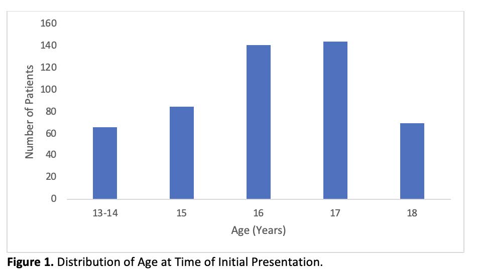
A total of 482 patients were included. Initial presentation of ACL injuries often occurred at 16 years old regardless of sport. Patients who were 13-14 or 18 years old (p = 0.009 and 0.035) or who received a tibialis anterior/bone-tendon-bone allograft (p = 0.002) were found to have increased risk of ACL re-tear. When adjusting for multiple variables, odds of ipsilateral re-tear in patients who received hamstring autograft (p = 0.02), sustained a contralateral ACL tear (p = 0.04), or a contact injury (p = 0.01) was increased.
Conclusions
High school freshman and seniors are found to have an increased risk of ACL re-tear. High school athletes must take caution when initiating sport participation (underclassman) and when playing at high intensity level (upperclassman), as susceptibility for ACL re-tear is elevated.
Can radiographic pre-planning, reduce the risk of tibial tuberosity fractures following osteotomy?
Jessica E. McCarthy, BVSc, MRCVS, DECVS
Assistant Professor in Small Animal Orthopedics, University of Wisconsin, Madison
Introduction and Purpose
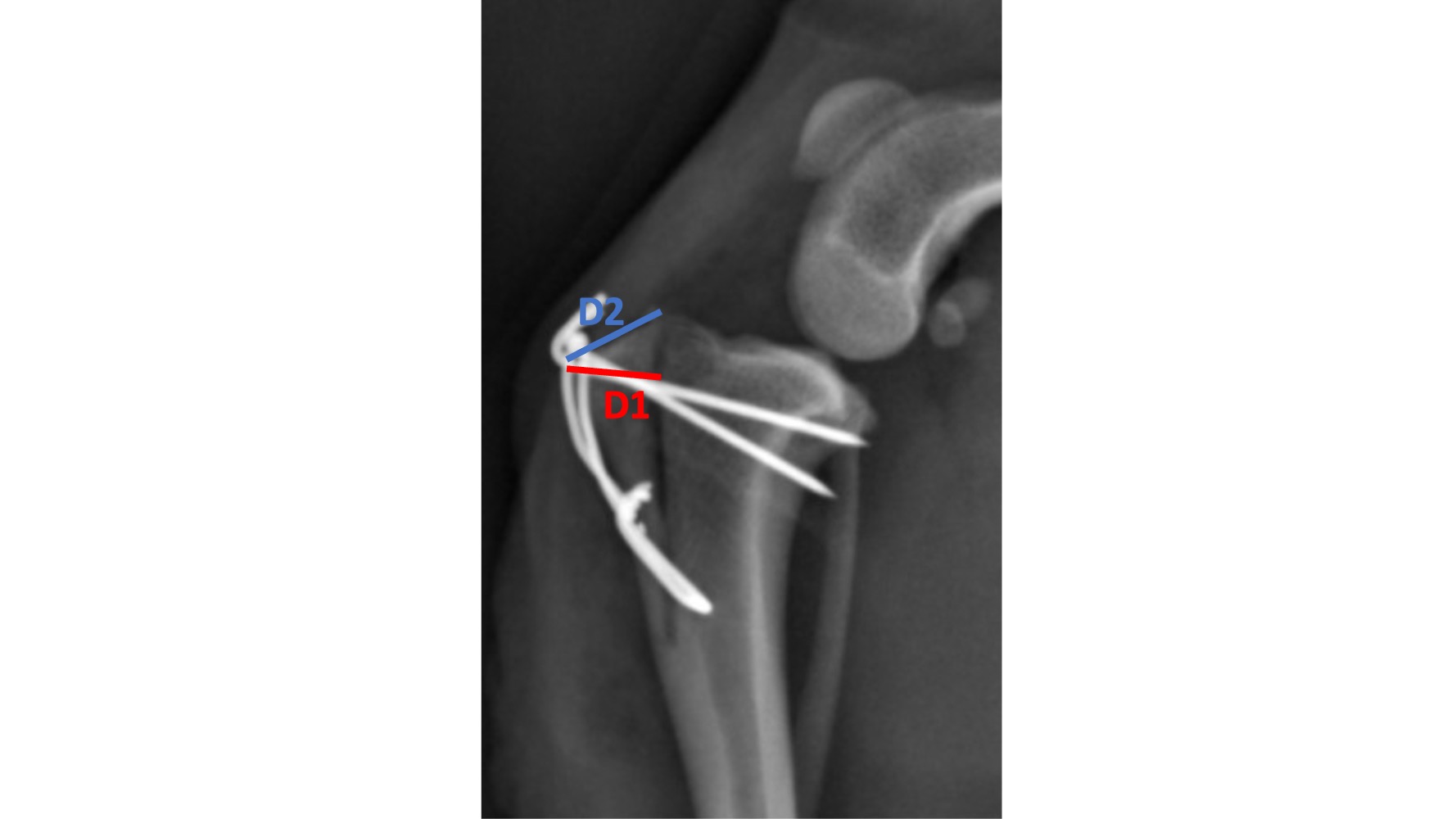
Tibial tuberosity osteotomy is often performed to treat patella luxation in dogs and to improve exposure for femoral osteotomy. Tibial tuberosity fracture occurs in up to 6% of cases. In patients undergoing tibial plateau leveling osteotomy (TPLO), there are measurement guidelines which reduce the risk of postoperative tibial tuberosity fracture. Our aim was to evaluate postoperative radiographs to identify whether these measurements, known as D1 and D2, can be used to plan tibial tuberosity osteotomies to reduce the risk of postoperative fracture.
Materials and Methods
Cases that had undergone surgical correction for patella luxation between 2000 and 2022 at three academic institutions were retrospectively identified. For radiographic measurements the medio-lateral radiographic view was used.
Results
267 dogs were identified, 50 dogs did not meet the inclusion criteria and were excluded giving a total of 217 dogs. Toy breeds were more likely to fracture than other breeds (p= 0.002). Pin position, size and number of pins did not affect the risk of tibial tuberosity fracture. A smaller (Log)D1 increased the risk of fracture (p=0.014). A D2 < 7mm had a sensitivity of 80% and specificity of80% for predicting dogs that will fracture.
Conclusions
Toy breeds should be considered at a higher risk of tibial tuberosity fracture than other breeds, and the smaller the size of the proximal tibia and tibial tuberosity segment, the greater risk. To reduce the risk of tibial tuberosity fracture, the value of D2 should be greater than, or equal to 7mm.
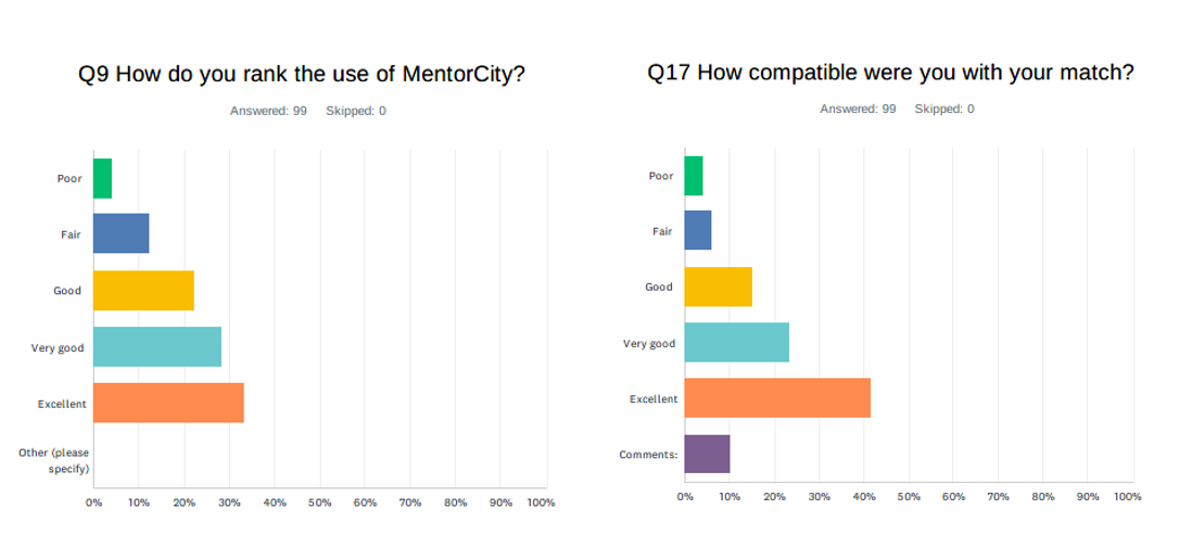
Formalized software-based mentorship pilot program - what has AO learned
Robert McGuire
AO Foundation
Introduction and Purpose
Mentorship works to challenge individuals’ perceptions of their potential career options, their roles within an organization, and the culture of their organization. Effective mentoring provides the tools to progress along chosen career paths as well as identifying future leaders. The purpose of the mentorship pilot was to a) measure the interest in a formalized software based mentorship program, b) test adaptability of an off-the shelf software solution and c) measure in interest in nonclinical competencies and across divisions.
Materials and Methods
Sourcing of software based mentorship program, creation of nonclinical competencies, open application for mentors and mentees, with anonymous matching, and a 3-months pilot program.
Results
Benefits and outcome
-
Widens access to mentorship
-
Supports unbiased mentorship (gender, ethnicity, region, etc)
-
Promotes significant professional, personal and social development (for mentor and mentee)
-
Creates a learning and leadership culture
-
Increases engagement and positively influences member satisfaction
-
Software proved to be successful
Conclusions
Launching a formalized mentorship program pilot - adapted with off-the-shelf software - as acritical next phase to create change at an individual and organizational level for a more diverse and inclusive organization. Supporting and creating different lived experiences through mentorship to enable a new future for AO members and faculty; where younger generations of surgeons, researchers and ORPs can share how to innovate, offering them opportunities that they have not had and allowing them to engage creating thriving communities while increasing our understanding our of diversity, so that everyone benefits from the change.
Biomechanical Analysis of a Modified Triangular Osteosynthesis Technique for Treatment of Spinopelvic Dissociation
Augustine M. Saiz, Jr., MD
Assistant Professor, UC Davis
Introduction and Purpose
Spinopelvic dissociation in young patients often consists of traumatic, high-energy bilateral sacral fractures. The purpose of this study is to biomechanically assess different fixation constructs for spinopelvic fractures to determine which construct provides the most stability.
Materials and Methods
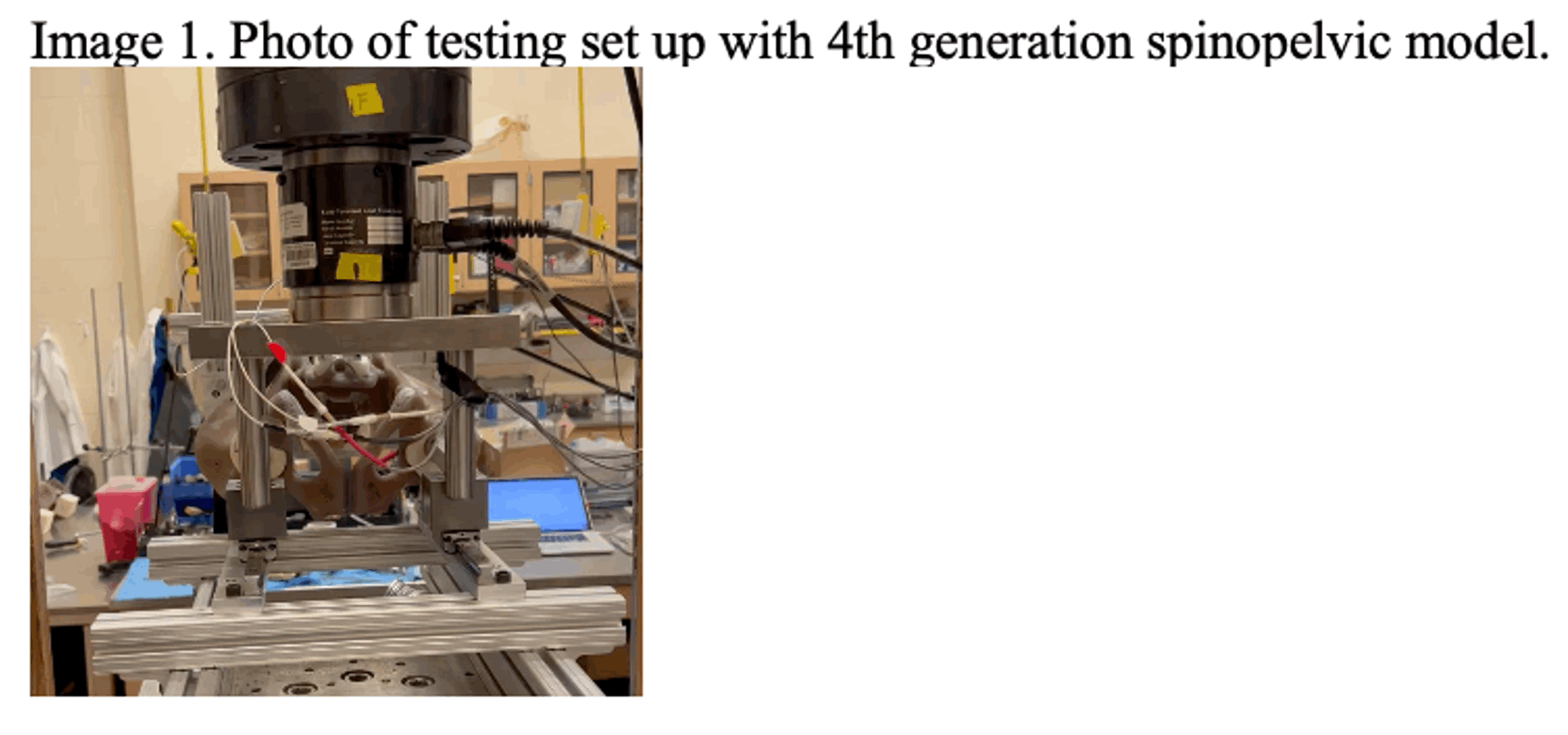
4th generation validated spinopelvic models composed of complete pelvises with L4 and L5vertebral bodies including ligamentous attachments were developed. A U-type sacral fracture with a
2 mm gap was created from the sacral ala through the S1 foramen with a transverse component atS1/S2 junction. 5 constructs in each model were tested: A) bilateral L5 pedicle screws with rods attached to iliac bolts (L5-IB), B) bilateral L5 pedicle screws with rods attached to iliac bolts with a transsacral-transiliac screw (L5-IB + TSTI), C) bilateral L5 and S1 pedicle screws with rods attached to iliac bolts with a transsacral-transiliac screw (L5-S1-IB + TSTI), D) bilateral L5 and S1pedicle screws with rods attached to iliac bolts (L5-S1-IB).
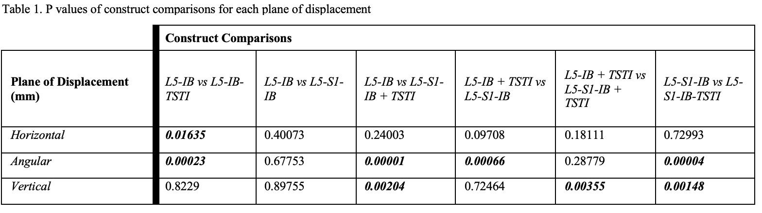
Results
The difference in horizontal, rotational, and vertical displacement between the L5-S1-IB + TSTI construct, and the other 3 constructs was significant (p < 0.05) with the L5-S1-IB + TSTI being the most stable. Furthermore, the L5-S1-IB + TSTI construct had the greatest load to failure vertically compared to the other 3 constructs (p < 0.05).
Conclusions
The modification of triangular osteosynthesis with S1 pedicle screws for lumbopelvic fixation in spinopelvic dissociation U-type fractures provides the greatest biomechanical fixation strength which may lead to less displacement and improved clinical outcomes.
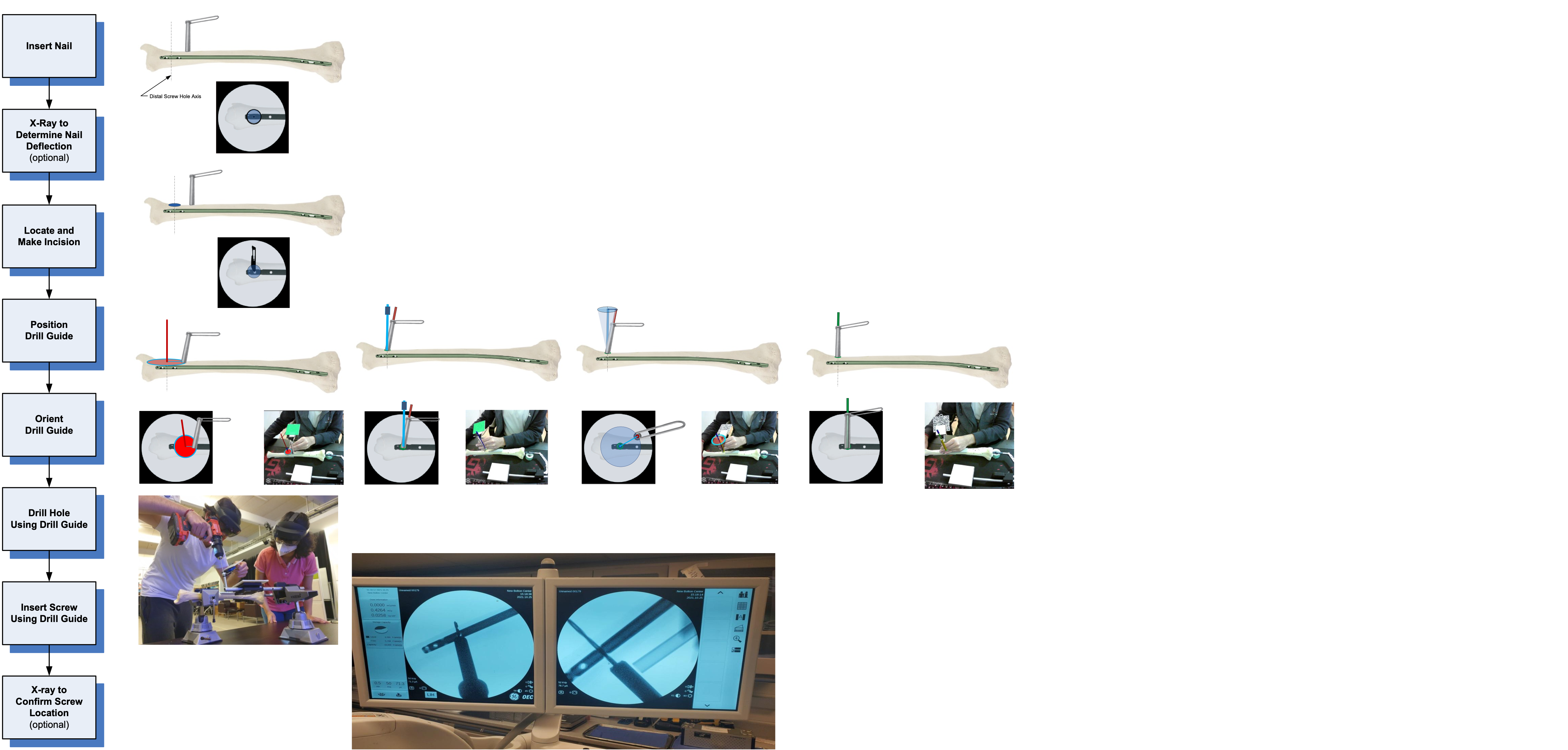
Augmented Reality Surgical Navigation System for Intramedullary Nailing, Remote Support & Surgical Training
Thomas P. Schaer, VMD
Dir Institute for Medical Translation New Bolton Center, University of Pennsylvania
Introduction and Purpose
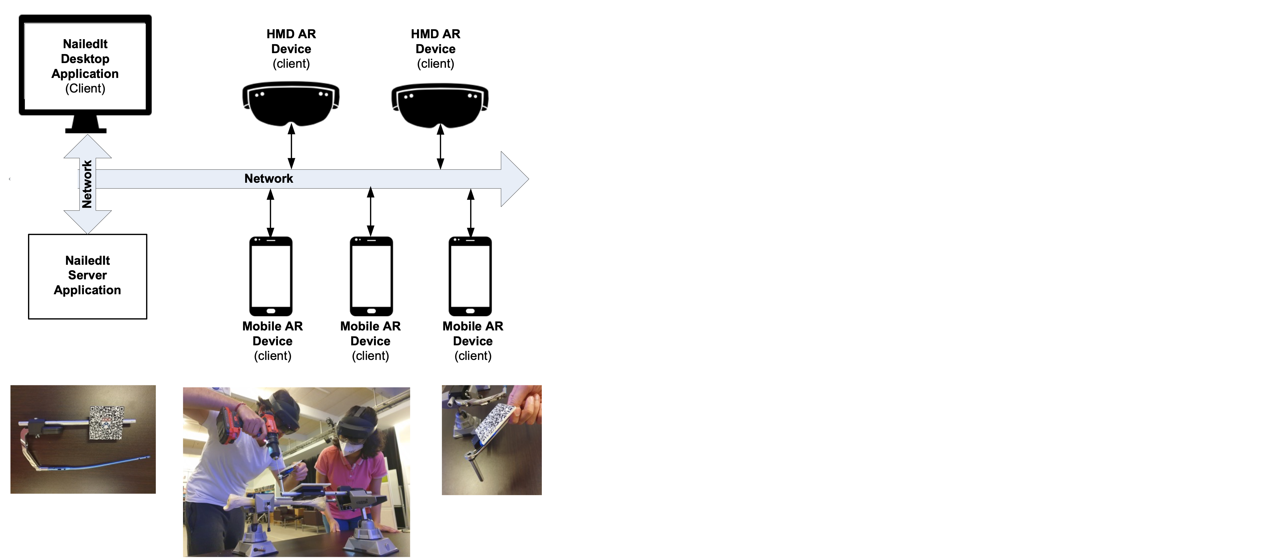
Intramedullary nailing (IMN) is considered the gold standard of long-bone fracture care. Accurate targeting of distal nail holes is the most difficult part. We developed a mixed-reality platform combining computer-assisted navigation, imaging targets on surgical instruments, and pertinent patient anatomy for tracking with augmented reality (AR) for IMN.
Materials and Methods
A customized registration app using HoloLens-to-world registration methods supported by external wireless inertial-measurement-unit orientation sensors and computer-vision image targets tracking both optical and fluoroscopic images is designed. During drilling, surgeons obtain accurate, in-situ visualization aids of the nail and the drilling path, and dynamic navigation is enabled.
Results
An intraoperative guidance system has been developed to provide intuitive visual and audio feedback cues of real-time deviations from the ideal path during the drilling procedure. Cadaver testing resulted in consistent and precise targeting when placing interlocking screws. Experiments showed reprojection errors along the X, Y, and Z axes were approximately 1.2 mm, 1.2 mm, and 1.2 mm, respectively. The end-to-end evaluation method indicated the distance error was 1.5 mm, and the 3D angle error was 2◦.
Conclusions
The augmented surgical navigation system provides surgeons with real-time guidance during orthopedic procedures eliminating time-consuming instrument registration or high-cost optical systems. Our navigation platform is agnostic of implant systems or manufacturers allowing for expansion beyond IMN. AR is assuming a fundamental and emerging role in surgical training and medical education. Its introduction results in providing learners with a better anatomic conceptualization and allows surgical simulations to improve their performances.
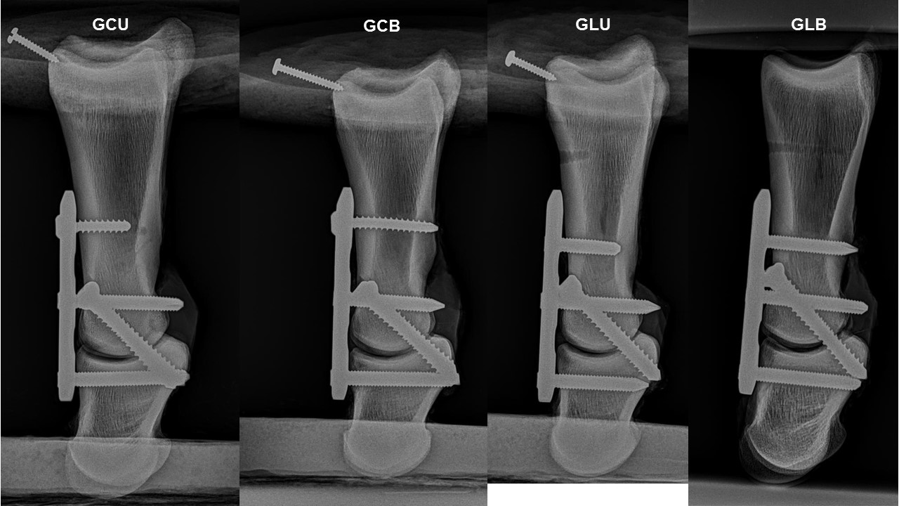
Biomechanical properties associated with different application forms of the proximal screw in equine pastern arthrodesis with locking compression plate
Anderson F. Souza
PhD student, Department of Surgery, School of Veterinary Medicine and Animal Science, University of São Paulo
Introduction and Purpose
The unicortical screw in the proximal hole of locking compression plate (LCP) has been recommended in pastern arthrodesis (PA) to avoid the occurrence of stress risers in the proximal phalanx. However, the cortical screws in this way do not follow the principles of internal fixation and may affect the stability of the construction. The objective of this study was to measure the biomechanical properties of PA with different forms of proximal screw application in LCP.
Materials and Methods
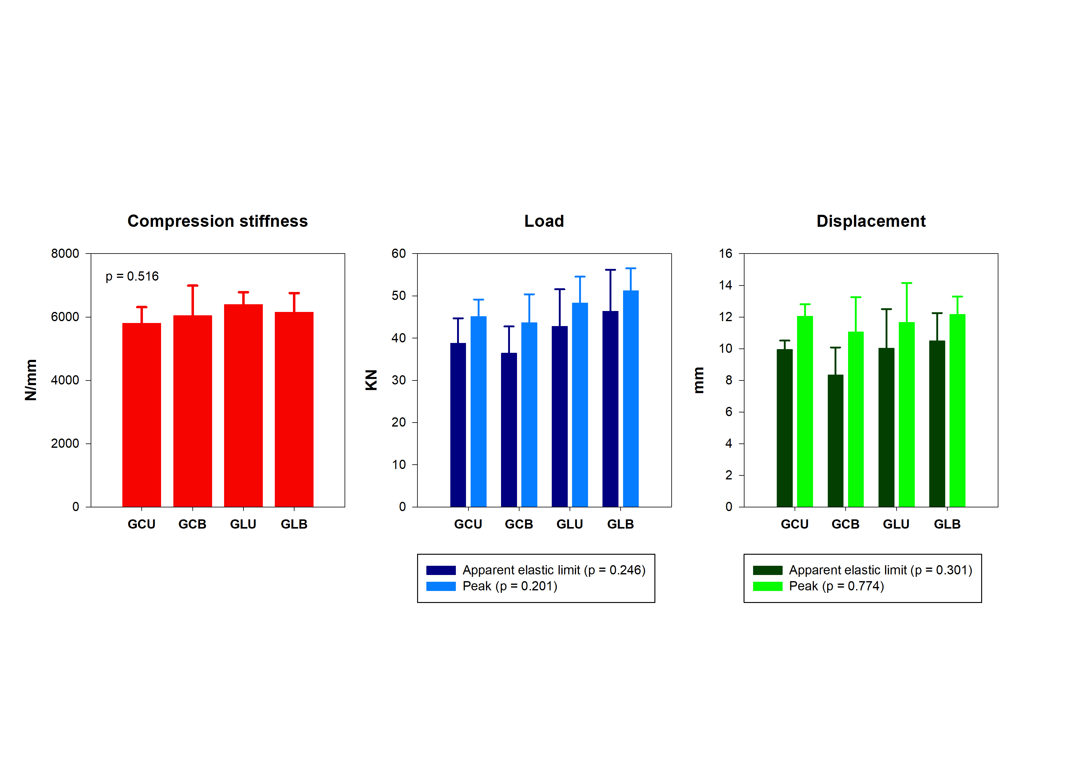
PA was performed in 20 cadaveric equine forelimbs, using a 3-hole 4.5mm LCP with two 5.5mmtransarticular cortical screws, divided into four groups according to the type of proximal screw applied to the plate: GCU - cortical screw unicortical; GCB - cortical screw bicortical; GLU -locking screw unicortical; and GLB - locking screw bicortical. A single-cycle axial compression testing to failure was performed. Compression stiffness (N/mm), load (N) and displacement (mm)at peak and apparent elastic limit were measured. Multivariate analysis of variance was used to compare the biomechanical variables between the groups, with the weight of the animals used as a cofactor (MANCOVA), followed by the post hoc Bonferroni test. P-values < 0.05 were considered significant.
Results
There were no significant differences between the biomechanical variables evaluated (P > 0.05).
Conclusions
The type of screw (locking or cortical) applied to the proximal hole of the locking plate and its form of anchorage (uni or bicortical) does not affect the biomechanical properties of equine pastern arthrodesis in cadavers submitted to single-cycle axial compression testing to failure.