Medical illustrator at work: putting Degenerative Cervical Myelopathy under the spotlight
"This project is a great example of how art can make science more accessible. In medicine, we often deal with concepts that cannot be seen with the naked eye. Even things that can be seen may be difficult to conceptualize. Because of this, diseases like Degenerative Cervical Myelopathy (DCM) benefit from some abstraction by an illustrator, to draw in the audience, educate them about the topic and entice them to ask more questions. That is the main goal of my work."
My name is Aaron Cole, and I am a medical illustrator at the Barrow Neurological Institute. I collaborated with Dr Rory Murphy and Dr Ben Davies to create an eye-catching illustration designed to shine a light on DCM. Our goal is to bring this poorly understood disease into the public eye in a different light. By raising awareness, patients can receive an earlier diagnosis and better prognosis. We hope that by fostering such educational outreach initiatives, we can resolve the Number 1 Research Priority of AO Spine RECODE-DCM: Raising Awareness.
I first learned about DCM while studying neuroanatomy and pathology in graduate school. Part of the discussion on the topic revolved around how little this disease is understood by the general public. This, along with the fact that DCM is likely to be underdiagnosed and has a wide array of symptoms, means that it is crucial to raise awareness so patients can receive a better prognosis.
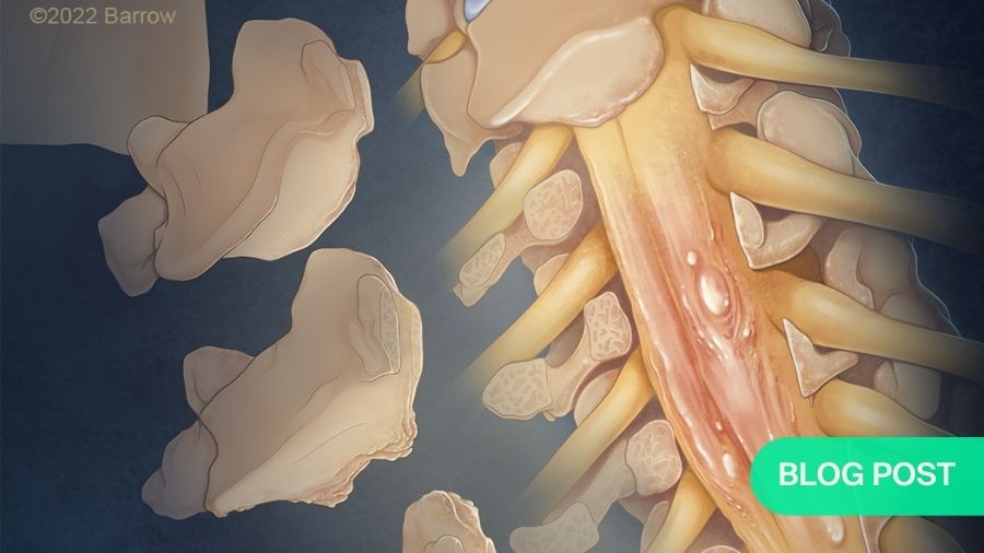
With Ben Davies, cofounder of Myelopathy.org, and Rory Murphy of the Barrow Neurological Institute, we wanted to create an eye-catching illustration that was designed to “shine a light” on DCM. This idea is credited to Murphy, who was interested in conceptualizing a way in which this poorly understood disease could be brought into the public eye.
It’s a little metaphorical and abstract in that sense, which I think shines through in the composition of the illustration.
All of my work begins with research, which usually revolves around reviewing the relevant anatomy and in this case learning how best to interpret the diseased portion of the vertebrae and spinal cord we wanted to show. From there, I’ll create a few rough drawings that try to encapsulate the tone and emotion of the project. This was very important for this project in particular, as most of my illustration work is very technical but here, we wanted to really home in on the feeling the illustration would convey.
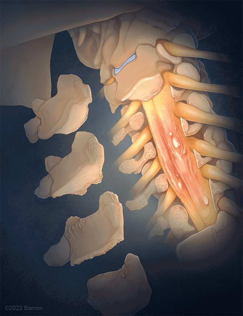
Once the final drawing was approved by Ben Davies and Rory Murphy, I began painting the final illustration using Adobe Photoshop. This is always the most fun part, as I can experiment with color, texture, lighting, and other elements to create something really eye-catching. To create the gif, I took the finished illustration and added some intense lighting effects in Adobe After Effects to give it the illusion of illuminating and coming to life.
I think this project is a great example of how art can make science more accessible, especially for a lay audience. In medicine we are often dealing with concepts that cannot be seen with the naked eye, and even things that can be seen are often difficult to conceptualize. Because of this, diseases like DCM benefit from some abstraction by an illustrator.
As you can see in this illustration, the goal is not to necessarily create something entirely accurate to human anatomy but to draw in your audience and educate them about the topic. Taking the liberty to remove structures or emphasize certain pathological portions of the anatomy results in something that not only captures the attention of the audience, but also entices them to ask more questions about what they’re looking at, and I would say that that is the main goal of my work.
About the author