

Critical clinical need for new approaches to promote intervertebral disc degeneration regeneration and recovery of function
BY DR ANDREA VERNENGO, DR ZHEN LI, AND DR SIBYLLE GRAD
Chronic low back pain (LBP), closely associated with intervertebral disc (IVD) degeneration, is a leading cause of disability worldwide and a significant burden for the health care and society. In the Disc Biology Special Issue of eCM Journal, we addressed the critical clinical need for new approaches that promote IVD regeneration and recovery of function.
The IVD is a complex organ consisting of multiple interrelated tissues. Cellular phenotype and function are unique with respect to cell development, mechanical environment, osmotic pressure, and hypoxia. The tissues are characterized by low cellularity, low nutrition, low turnover and avascularity, posing a significant challenge to researchers aiming to repair them. Towards this goal, a detailed understanding of the IVD biochemistry is crucial for finding effective targets that maintain the homeostasis and prevent, decelerate, or reverse adverse signaling. Recent research has identified many underlying molecular signals and pathways that maintain the physiological state in a healthy IVD. This knowledge allows, in turn, for the detection of aberrant regulations that may lead to cell and matrix deterioration and, finally, to the destabilization of the spinal structure.
In this Special Issue, different molecular markers and signaling pathways, potentially contributing to the maintenance or disturbance of the IVD homeostasis, were described. Li et al. (2020) identified the existence of the tissue renin-angiotensin system (tRAS) in human IVD tissue. The results suggested a correlation between tRAS expression, inflammation and IVD degeneration. Snuggs et al. (2021) found that aquaporin 1 and 4 play important roles in the fundamental adaptation of nucleus pulposus (NP) cells to their osmotic environment. The function of these proteins may be lost during disc degeneration, when aquaporin 1 and 4 expression is reduced. Hayes and Melrose (2021) studied the three-dimensional distribution of perlecan within IVD chondrons. The results suggested the possibility of the involvement of this heparan sulphate proteoglycan in an intracrine regulatory system and its potential as a novel therapeutic target. Joyce et al. (2021) investigated the role of altered glycosylation in human NP cells under inflammation-induced degeneration condition and identified potential therapeutic targets for future regenerative therapies. Toll-like receptor (TLR) signaling results in the release of pro-inflammatory cytokines and proteolytic enzymes that can directly cause IVD degeneration and back pain. Bisson et al. (2021) provided a timely review on TLR activation in IVD degeneration and discussed potential treatment modalities to alleviate the inflammatory phenotype of disc cells to arrest IVD degeneration and back pain.
To counteract the deteriorating cascades at an early stage, various cellular and molecular therapies are reported that hold great promise for mitigating or reversing IVD degeneration. Panebianco et al. (2020) reviewed the state of the art in injectable biomaterial cell carriers, highlighting the important need for next-generation biomaterials that can provide both significant initial biomechanical restoration and a functional niche permitting biological function of delivered cells. Gansau and Buckley (2021) studied the important challenge of injected cell survival in the acidic microenvironment of the degenerated IVD. They showed that priming bone-marrow-derived stem cells (BMSCs) and articular chondrocytes (ACs) with transforming growth factor (TGF)-β3 mitigated the detrimental effects of low pH on cell viability and matrix accumulation. Jacobsen et al. (2021) addressed the issue of inflammation, an important factor that accelerates IVD matrix loss during degeneration and can hamper the effectiveness of cell-based repair strategies. Their study demonstrated that inhibition of TLR4 can block pro-inflammatory signalling and degenerative changes to NP cell biophysical properties. Tang et al. (2021) presented the delivery of engineered vesicles (EVs) loaded with forkhead-box F1 (FOXF1) mRNA as a minimally invasive, non-viral and clinically feasible approach for re-programming adult IVD cells towards healthier phenotypes.
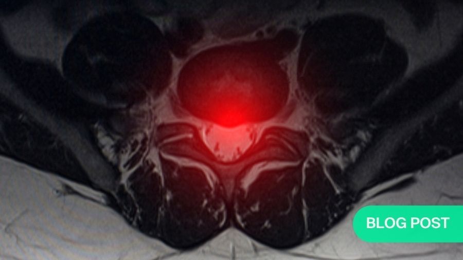
The function of the IVD strongly depends on its mechanical integrity. In this context, Wade et al. (2020) reported on the impact of pre-existing annular defects on the susceptibility to disc herniation upon complex mechanical load. Using motion segments from mature sheep, the authors found a clear association between the presence of structural changes in the inner and mid annulus fibrosus (AF) and the occurrence of NP displacement or, eventually, disc herniation following overloading. This finding improves the knowledge on the role of mechanical load in the development of IVD degeneration and the mechanism of trauma-induced disc herniation. Furthermore, the work demonstrates the use of ultra-high field magnetic resonance imaging (MRI) to detect discs at risk of herniation.
The comprehensive review article by Ashinsky et al. (2021) highlighted the broader concept of a whole motion segment approach to study degeneration and design regenerative therapies. Specifically, the interfaces between NP/AF, cartilaginous endplate, bony endplate, and vertebral bone may experience crucial changes during degeneration. When characterising these interfaces, it is also important to consider the vascular morphology within the bony endplate. Finally, the contribution of the facet joints and spinal muscles to pain and degeneration are still ill defined. Animal models or advanced whole-organ culture and co-culture models, using human tissues, are warranted to study the crosstalk between the spinal tissues in health and disease.
Preclinical research is strongly directed by the specific study aim and involves both in vitro and in vivo evidence. Mouse models are instrumental for fundamental studies on molecular mechanisms. A comprehensive overview of genetic mouse models and their characteristics was presented by Melrose et al. (2021). Despite the distinct differences in development and structure between the murine and human spine, mouse models have proven extremely valuable in the elucidation of developmental signals and pathological pathways, while translation of the findings to larger animals and ultimately to the human patient will be the next steps in research. Kim et al. (2021) investigated the expression of the mechano-sensitive ion channel transient receptor potential vanilloid 4 (TRPV4) during mouse spinal development and postnatal maturation as well as after mechanical stimulation of AF cells. The distinct spatio-temporal expression during IVD development and the TRPV4-mediated regulation of matrix gene expression after mechanical stimulation indicate a prominent mechano-biological role of this ion channel. Another developmental study, using muscular dysgenesis mice, was performed by Levillain et al. (2021). At prenatal stages, the influence of muscular forces on the development of the vertebrae and the IVD was investigated. In the absence of muscle contractions, the authors observed vertebral fusions, NP displacement and aberrant development of the AF, including disrupted collagen fibers, which emphasizes the importance of mechanical forces for functional AF tissue formation.
With respect to large animal models, the sheep has been one of the most widely used preclinical in vivo models for IVD degeneration and repair. While degeneration is typically induced by mechanical or biochemical means, Bouhsina et al. (2021) characterized the natural ageing of the ovine lumbar IVD. Imaging and histological results confirmed the development of age-related disc degeneration in the ovine lumbar IVDs; hence, the ovine species, showing a similar IVD structure to that found in human spine, could be valuable to assess regenerative therapies without the need to induce degeneration by external means.
Taken together, complementary efforts of preclinical and clinical research are indispensable for a precise definition of biological and biomechanical needs and their reliable relation to the clinical needs. The developments presented herein represent a significant contribution towards the overall aim of improved patient care.
All published articles of the Disc Biology Special Issue can be accessed here.
About the authors

Andrea Vernengo, Assoc Prof, PhD, is a researcher in the Regenerative Orthopaedics Program at the AO Research Institute Davos (ARI) pursuing research projects related to the interverbal disc, including induced pluripotent stem cell-based regeneration strategies and 3D bioprinting of cell-instructive scaffolds for annulus fibrosus tissue engineering.


References and further reading:
- Ashinsky B, Smith HE, Mauck RL, Gullbrand SE (2021) Intervertebral disc degeneration and regeneration: a motion segment perspective. Eur Cell Mater 41: 370-380.
- Bisson DG, Mannarino M, Racine R, Haglund L (2021) For whom the disc tolls: intervertebral disc degeneration, back pain and toll-like receptors. Eur Cell Mater 41: 355-369.
- Bouhsina N, Decante C, Hardel JB, Madec S, Abadie J, Hamel A, Le Visage C, Lesoeur J, Guicheux J, Clouet J, Fusellier M (2021) Correlation between magnetic resonance, X-ray imaging alterations and histological changes in an ovine model of age-related disc degeneration. Eur Cell Mater 41: 166-178.
- Gansau J, Buckley CT (2021) Priming as a strategy to overcome detrimental pH effects on cells for intervertebral disc regeneration. Eur Cell Mater 41: 153-169. Hayes AJ, Melrose J (2021) 3D distribution of perlecan within intervertebral disc chondrons suggests novel regulatory roles for this multifunctional modular heparan sulphate proteoglycan. Eur Cell Mater 41: 73-89. Jacobsen TD, Hernandez PA, Chahine NO (2021) Inhibition of toll-like receptor 4 protects against inflammation-induced mechanobiological alterations to intervertebral disc cells. Eur Cell Mater 41: 576-591.
- Joyce K, Mohd Isa IL, Krouwels A, Creemers L, Devitt A, Pandit A (2021) The role of altered glycosylation in human nucleus pulposus cells in inflammation and degeneration. Eur Cell Mater 41: 401-420.
- Kim MK, Ramachandran R, Seguin CA (2021) Spatiotemporal and functional characterisation of transient receptor potential vanilloid 4 (TRPV4) in the murine intervertebral disc. Eur Cell Mater 41: 194-203.
- Levillain A, Ahmed S, Kaimaki DM, Schuler S, Barros S, Labonte D, Iatridis JC, Nowlan NC (2021) Prenatal muscle forces are necessary for vertebral segmentation and disc structure, but not for notochord involution in mice. Eur Cell Mater 41: 558-575.
- Li Z, Wystrach L, Bernstein A, Grad S, Alini M, Richards RG, Kubosch D, Sudkamp N, Izadpanah K, Kubosch EJ, Lang G (2020) The tissue-reninangiotensin-system of the human intervertebral disc. Eur Cell Mater 40: 115-132.
Melrose J, Tessier S, Risbud MV (2021) Genetic murine models of spinal development and degeneration provide valuable insights into intervertebral disc pathobiology. Eur Cell Mater 41: 52-72. - Panebianco CJ, Meyers JH, Gansau J, Hom WW, Iatridis JC (2020) Balancing biological and biomechanical performance in intervertebral disc repair: a systematic review of injectable cell delivery biomaterials. Eur Cell Mater 40: 239-258.
- Snuggs JW, Bunning RA, Le Maitre CL (2021) Osmotic adaptation of nucleus pulposus cells: the role of aquaporin 1, aquaporin 4 and transient receptor potential vanilloid 4. Eur Cell Mater 41: 121-141.
- Tang S, Salazar-Puerta A, Richards J, Khan S, Hoyland JA, Gallego-Perez D, Walter B, Higuita-Castro N, Purmessur D (2021) Non-viral reprogramming of human nucleus pulposus cells with FOXF1 via extracellular vesicle delivery: an in vitro and in vivo study. Eur Cell Mater 41: 90-107.
Wade K, Berger-Roscher N, Rasche V, Wilke H (2020) Disc wall structural abnormalities can act as initiation sites for herniation. Eur Cell Mater 40: 227-238.
Disclaimer
The articles included in the AO Spine Blog represent the opinion of individual authors exclusively and not necessarily the opinion of AO Spine or AO Foundation.