Degenerative Spondylolisthesis and Retrolisthesis - relationship with whole body radiographic parameters
BY HEY HWEE WENG DENNIS, TAN JUN HAO, TAN TUAN HAO
In the landscape of adult spinal deformity, understanding the intricate relationship of spondylolisthesis and retrolisthesis with whole-body radiographic parameters is the key to determining the ideal surgical treatment for these conditions.
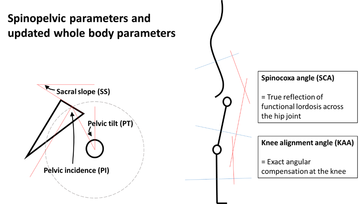
The rapidly evolving concepts in adult spinal deformity surgery have highlighted the importance of optimal sagittal spinal profile in achieving good outcomes. This entails a meticulous clinical and radiographic evaluation, incorporating metrics such as the sagittal vertical axis (SVA), pelvic incidence-lumbar lordosis (PI-LL) discrepancy, and the pelvic tilt (PT) – a radiographic marker for assessing compensatory mechanisms at the hip for positive sagittal imbalance. New parameters such as the knee alignment angle (KAA) and spinocoxa angle (SCA) are equally important to investigate into the behaviour of whole body balance.
Focally, retrolisthesis which was once regarded as a possible form of spinal instability of unknown significance, is now being increasingly recognized today as possibly yet another compensatory process which occurs in patients with positive sagittal imbalance.
Using updated whole-body radiographic parameters measured by the slot scanner, we attempted to understand the association between these parameters, hence hypothesizing the pathoaetiology of various forms of spondylolisthesis and retrolisthesis. This study was awarded one of the two best papers of 2023 in Global Spine Journal.
Understanding spondylolisthesis and retrolisthesis in the context of whole body balancing considering postural variations
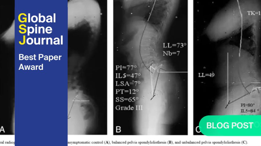
Spondylolisthesis, the forward translation of the vertebra body over its caudal counterpart, is more prevalent in patients above 50 years old, and is likely reflective of degenerative aetiology and exhausted physiological reserves with acute worsening of the condition. This is supported by the concomitant findings of sagittal compensation such as increased PT, KAA and SCA in patients with spondylolisthesis (Figure 1).
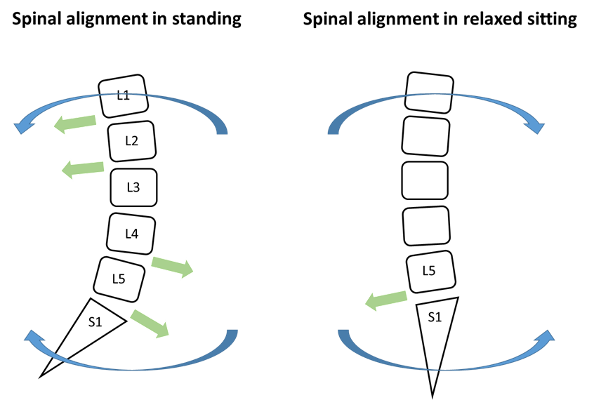
On the other hand, retrolisthesis, backward translation of the vertebra body over its caudal counterpart, is seen in all age groups and increases gradually with age, suggesting its possible compensatory role in sagittal balancing rather than being the primary pathology. Unlike spondylolisthesis which is often a pathological problem resulting from shear forces residing in the lordotic lower lumbar spine, as well as during forward bending postures and activities, primary retrolisthesis is less common as minimal postures or activities drive the spine backwards. Nevertheless, Hey et al (2022) has shown that degenerative retrolisthesis and loss of lordosis at L5-S1, which is a common phenomenon, is likely a result from long-standing lower lumbar spine bending forces against the posterior ligamentous complex during slump sitting, predisposed by a negatively sloped sacrum and increased lumbar flexibility (Figure 2).
Increased thoracolumbar junctional angle (TLA) which was associated with retrolisthesis are again suggestive of adaptive changes following prolonged natural standing or sitting postures where the patient employs posterior ligamentous complex at the thoracolumbar spine to balance and conserve energy. This shifts the body’s centre of gravity backwards likely resulting in a backward shear force on the thoracolumbar and upper lumber vertebrae.
While it is possible that both spondylolisthesis and retrolisthesis vary somewhat in terms of aetiology, the result of vertebra translation should depend on the similar force principles. A forward-directed nett force should lead to spondylolisthesis and a backward-directed nett force retrolisthesis. Determinants of these forces include the posture and activities of the patient, which in turn influences the force magnitude on each lumbar vertebra. In our study population, the higher prevalence of spondylolisthesis from L3/4 to L5/S1 levels and retrolisthesis from L1/2 to L3/4 further substantiates this point. Since lordosis occurs in the lumbar spine particularly the lower segments, and lower segments have a positive slope angle, under the pull of gravity, forward vertebral translation naturally ensues. Cranial to the apex of the lumbar curve, which usually resides in L3/4, an opposite force vector results in backward vertebral translation (Figure 2).
Spondylolisthesis and retrolisthesis in association with spinopelvic slope angles
In spondylolisthesis patients, higher PI values was noted compared to those without. This observation highlights the role of individual-specific pelvic morphology as a risk factor for the development of spondylolisthesis.
Additionally, higher PT and SS, alongside changes in LL and SCA were evident in association with spondylolisthesis. While PT can fluctuate with postural changes and is influenced by spinopelvic compensation mechanisms, the simultaneous increase in SS, which conventionally acts reciprocally with PT, reinforces that this increase is likely to be also contributed by pelvic morphology. This can be further explained by the equation PI = PT + SS, where patients with a large PI naturally bears a larger PT and SS. This hypothesis is further supported in the observation of larger L5I, L5T and L5S associated with L4/5 spondylolisthesis. The absence of similar findings in other intervertebral levels is likely due to the low prevalence of spondylolisthesis at those levels.
In the pathogenesis of spondylolisthesis, both the prevailing clinical literature and our study findings highlight the presence of larger L4I and L4S as indicative of a significant association with L3/4 spondylolisthesis.
Notably, although there is a tendency for sacral slope to predict L5/S1 spondylolisthesis, this finding might be confounded by the intrinsic rigidity at the L5/S1 level that certain patients experience due to transitional vertebrae and iliolumbar ligament involvement.
The presence of retrolisthesis in association with a smaller L4S, as well as a smaller SS is interesting and may be due to postural changes in sitting that stresses the spine in pelvic retroversion discussed earlier. This phenomenon may stem from postural dynamics, particularly in seated positions, which exert stress on the spine through pelvic retroversion.
Future direction
Surgical treatment of spinal conditions depends greatly on differentiating between pathology and physiology - the former must be rectified for symptom relief while the latter accommodated. With failure of non-operative treatment, spondylolisthesis which is often pathological, must be treated. Physiological compensatory mechanisms of the spine such as retrolisthesis and whole body adjustments can turn into pathological diseases with neglect, and has to be respected.
Our work provides a new understanding of the intricate dynamics between spondylolisthesis, retrolisthesis and whole body balance. Moving forward, larger-scale studies hold greater promise in unravelling more intricate dynamics between these conditions, thereby improving our capabilities in treating these conditions.
References and further reading:
- , , , , , , , , , . Prevalence and Risk Factors of Degenerative Spondylolisthesis and Retrolisthesis in the Thoracolumbar and Lumbar Spine - An EOS Study Using Updated Radiographic Parameters. Global Spine J. 2024 May;14(4):1137-1147.doi:10.1177/21925682221134044
- , , , , . Understanding the Pathophysiology of L5-S1 Loss of Lordosis and Retrolisthesis: An EOS Study of Lumbopelvic Movement Between Standing and Slump Sitting Postures. World Neurosurg 2022 Feb:158:e654-e661.doi: 10.1016/j.wneu.2021.11.034.
About the authors:
Hey Hwee Weng Dennis is a senior consultant (attending) spine surgeon currently working in the Department of Orthopaedic Surgery, National University Health System, Singapore. He is also an assistant professor at the National University of Singapore. He has a practice heavy on adult spinal deformity, which he treats increasingly with artificial disc replacements and endoscopic spine surgeries, on top of conventional spinal instrumented fusion surgeries. His passion to constantly improve his patient’s outcomes led him to develop a novel and comprehensive patient-specific, spinal alignment assessment radiographic protocol that respects multiple spinal postures (in standing and in sitting) to aid surgical planning, and to establish the Motion-preserving and Minimally-invasive Spine Unit (MMSU) in his hospital – a unit that focuses on preserving/restoring physiology whilst treating the highly complex condition, that is adult spinal deformity.
You can find out more about his past and ongoing projects (here) https://www.nuh.com.sg/patients-visitors/Pages/find-a-doctor-details.aspx?docid=Hey_Hwee_Weng_Dennis
You might also be interested in:
AO Spine Knowledge Forums (KF)
Creating new knowledge to make your patients and practice flourish.
Courses and events
Find all upcoming courses and events on lumbar pathologies and approaches.
Global Spine Journal
AO Spine’s official scientific journal, the first fully open access journal in the field of spine surgery with an Impact Factor.


