
Orthopedic challenges in miniature and toy-breed dogs
BY DR TARO KIMURA

Orthopedic disease in miniature and toy-breed dogs is an important part of modern small animal orthopedic practice. In many regions, including large parts of Asia, these dogs now represent a substantial proportion of the cases referred for surgical management. While the diagnoses may appear familiar, the underlying biomechanics, anatomical constraints, and surgical risks are often very different from those encountered in larger breeds. I have come to view these cases not as “small versions” of large-breed problems, but as a distinct orthopedic category that demands its own mindset, planning strategy, and technical discipline.
-
Read the quick summary:
- Dr Taro Kimura discusses orthopedic disease in miniature and toy-breed dogs, focusing on MPL, CCLD, limb deformities, and the constraints of small skeletal anatomy.
- His focus is on the need for limb-wide assessment, precise deformity correction, and strict application of AO principles to achieve durable outcomes.
- Practical guidance on planning, alignment analysis, and adapting fixation and osteotomy strategies to miniature patients is included.
- Further discussion centers on how extensively advanced imaging and corrective surgery should be applied in routine versus complex small-breed cases.
Disclaimer: The article represents the opinion of individual authors exclusively and not necessarily the opinion of AO or its clinical specialties.
Developmental disorders and the problem of isolated treatment
Developmental conditions dominate the caseload. Medial patellar luxation (MPL), hip dysplasia (HD), and cranial cruciate ligament disease (CCLD) account for most presentations, frequently occurring in combination rather than isolation. In miniature dogs, abnormalities of the quadriceps–patella–tibial mechanism are often accompanied by femoral varus, tibial torsion, and altered joint orientation. Treating a single joint without addressing global limb alignment rarely produces durable results. These patients challenge us to step back, assess the entire limb, and respect the cumulative effect of multiple small deformities.
Scale is a defining feature of miniature-breed orthopedics. Small bones leave little room for error, whether in implant placement, osteotomy execution, or alignment correction. A few degrees of residual varus or torsion may be clinically irrelevant in a large dog, yet biomechanically disastrous in a two kilogram patient. Implant tolerance is narrow, soft tissues are unforgiving, and fixation failure can occur with even subtle technical inaccuracies. This forces a higher level of discipline in preoperative planning and intraoperative execution.
Medial patellar luxation as a limb-wide disease
Medial patellar luxation illustrates these challenges particularly well. In many small-breed dogs, MPL is not a disease of the patella itself but the visible endpoint of a complex developmental process. Femoral varus, distal femoral procurvatum, internal tibial torsion, medial displacement of the tibial tuberosity, and trochlear dysplasia frequently coexist. Correcting only the patellar tracking without addressing underlying deformities may temporarily improve gait but leaves abnormal load transmission unchanged. In our experience, recurrence and progression of degenerative joint disease are often the consequences.
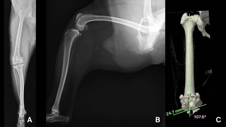
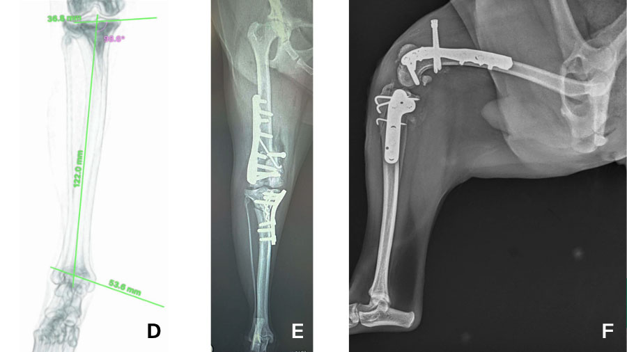
Figure 1. Preoperative radiographs and computed tomography (CT) images of the right pelvic limb in a Japanese Chin with grade II medial patellar luxation presenting with intermittent lameness. (A–B) Preoperative craniocaudal and mediolateral radiographs demonstrating abnormal alignment of the distal femur and tibia, with osteophyte formation of the femoral trochlea. (C) Three-dimensional CT reconstruction demonstrating distal femoral varus (aLDFA = 107°) and external femoral torsion. (D) Frontal-plane CT reconstruction of the tibia demonstrating external tibial torsion. (E) Postoperative craniocaudal radiograph demonstrating medial translation of the distal femoral fragment to correct the femoral deformity while preserving the mechanical axis in accordance with Paley's principles. (F) Postoperative mediolateral radiograph demonstrating the distal femoral osteotomy stabilized with plate fixation. An antirotational screw was placed to maintain alignment of the extensor mechanism and prevent medial displacement of the quadriceps mechanism. A tibial plateau leveling osteotomy (TPLO) was also performed to treat the concurrent cranial cruciate ligament rupture (CCLR).
Cranial cruciate ligament disease in abnormal stifles
Cranial cruciate ligament disease adds another layer of complexity. In miniature breeds, CCLD often develops in an already abnormal stifle. Distal femoral varus, abnormal tibial plateau geometry, and concurrent MPL alter normal kinematics long before the ligament fails. Instability patterns may differ from those described in larger dogs, and standard extracapsular or osteotomy-based techniques may not adequately address the true biomechanical problem. These cases remind us that ligament rupture is frequently a symptom rather than the primary disease.
For this reason, I approach cruciate disease in small dogs with a strong emphasis on alignment analysis. Evaluating frontal, sagittal, and axial plane deformities is critical before selecting a surgical strategy. In some patients, addressing femoral or tibial deformity is as important as stabilizing the stifle itself. Modified osteotomy techniques, combined procedures, or staged corrections may be required to restore functional biomechanics. Although these approaches are more demanding, they offer the best chance for predictable long-term outcomes.
Advanced imaging and surgical planning
Advances in diagnostic imaging and surgical planning have significantly expanded what is achievable in miniature dogs. High-resolution computed tomography allows accurate assessment of torsion, joint orientation, and multiplanar deformity that cannot be reliably evaluated on radiographs alone. Digital templating and three-dimensional planning facilitate precise osteotomy design and implant selection, which is particularly valuable when bone stock is limited. Patient-specific instrumentation and cutting guides can further reduce intraoperative variability and improve accuracy.
These tools have transformed the management of conditions that were previously considered extremely challenging or even untreatable. Genu recurvatum, congenital elbow luxation, and complex multiplanar limb deformities can now be addressed more predictably using techniques adapted to miniature skeletal anatomy.
However, technology should support, but not replace, clinical judgment. Planning software cannot compensate for a poor understanding of biomechanics, nor can it override the need for meticulous surgical technique.
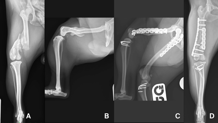
Figure 2. Preoperative and postoperative radiographs of distal femoral malunion in a Pug. (A–B) Preoperative craniocaudal and mediolateral radiographs demonstrating severe distal femoral malunion with multiplanar angular deformity. (C–D) Postoperative mediolateral and craniocaudal radiographs confirming restoration of femoral alignment following corrective osteotomy and stabilization using orthogonal locking plates.
A balanced approach for the AO surgeon
Successful outcomes in miniature-breed orthopedics remain grounded in fundamental AO principles. Respect for soft-tissue biology is paramount, as vascular compromise or excessive dissection can quickly lead to delayed healing or implant failure. Mechanical alignment must be restored with the same rigor applied in larger patients, if not more so. Fixation must be stable yet appropriate to bone quality, avoiding oversized implants that risk stress shielding or iatrogenic fracture.
Load distribution deserves particular attention. In small dogs, forces are concentrated over a limited bone surface, and implants are often subjected to proportionally high stresses. Thoughtful implant selection, accurate placement, and careful postoperative management are essential. I am increasingly convinced that many complications in miniature-breed surgery stem not from disease severity, but from underestimating the biomechanical consequences of minor technical errors.
These cases have reshaped how I counsel owners. I emphasize that orthopedic disease in miniature dogs is often complex, multifactorial, and developmental in nature. Surgical success depends on addressing the true underlying problem rather than the most obvious clinical sign. While advanced planning and technology may increase upfront cost, they can often reduce the risk of revision surgery and improve functional longevity.
For surgeons within the AO network, miniature and toy-breed dogs offer an opportunity to apply core principles at the highest level of precision. These patients demand careful assessment, disciplined planning, and technical excellence. When advanced tools are integrated thoughtfully and AO principles are applied without compromise, reliable outcomes are achievable even in cases that once seemed beyond reach. In our view, this balanced approach represents the future of small-breed orthopedic surgery.
About the authors:

Dr Taro Kimura, DVM, PhD, is a veterinary orthopedic surgeon based in Tokyo, Japan, with extensive clinical, academic, and teaching experience in small animal surgery. Born and raised in a veterinary hospital, Dr. Kimura developed an early understanding of the realities of animal disease, pain, and clinical responsibility.
At the beginning of his professional career, Kimura trained in veterinary anesthesia, establishing a solid foundation for safe, consistent, and reliable surgical care through rigorous perioperative management and risk control. Guided by a desire to relieve pain, restore mobility, and preserve each animal’s quality of life, he subsequently transitioned into orthopedic surgery, with a particular focus on joint replacement surgery and the surgical management of complex orthopedic conditions, including congenital and difficult-to-treat disorders.
Dr Kimura is the Director of Vet Surg Tokyo (VST), a private referral orthopedic practice, while also serving as Director of Kimura Animal Hospital. In parallel with his clinical work, he has contributed to veterinary education as an Assistant Professor at Tokyo Medical University and Yamazaki Professional College of Animal Health Technology.
His clinical experience in joint replacement surgery, complex fracture management, and advanced orthopedic procedures has been reported in peer-reviewed international journals. Dr Kimura is committed to applying insights gained from daily clinical practice to subsequent patient care, emphasizing careful decision-making, surgical safety, and reproducibility.
You might also be interested in...

Small Animal Fractures
Principles of fracture management in small animals: assessment, planning, and performance.
AO Surgery Reference
An internet-based resource for the management of equine, dog, and cat fractures.

AO VET Education
Delivering life-long education through courses and training worldwide.
Clinical library
Explore our educational resources, develop your competencies, and improve patient care.