
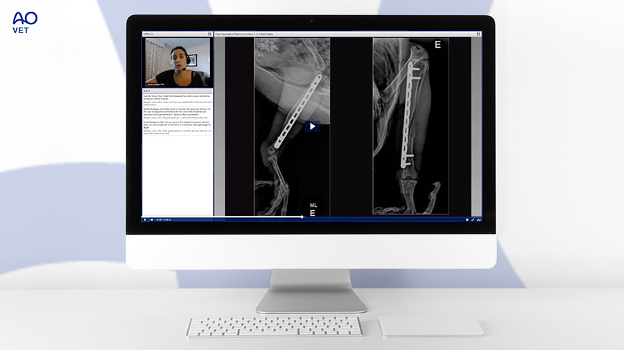
AO VET Videos and webinars
Discover the full collection of educational videos with small and large animal practical exercises, recorded webinars and expert presentations by AO VET's world-renowned faculty. AO VET Members have unlimited access to all video materials, AO TV videos are open to all registered users.
Large animals
2023
Meet the Experts - Veterinary Large Animal Screw Targeting Clamp
Proximal Phalangeal Fracture in Equine
2019
Racehorse Injuries: Fact Vs. Fiction—Where Do AO VET Surgeons Fit In
2011
Core Decompression of the Equine Navicular Bone
2010
AO VET Equine Course - Arthroscopically Assisted Fracture Repair
Small animals
2020
Orthopedic Challenges in Sport Dogs
2019
Discussion of Controversial Topics in Veterinary Medicine
2010
AO VET Small Animal - Implant Failure
AO VET Small Animal - Nonunions: The Simple, The Bad and The Ugly
Large animals
2023
Appropriate Treatment of Distal Phalanx Fractures in Horses
Arthroscopy in the Horse—Challenging Cases and their Solutions (Spanish)
Cómo Sacar El Máximo Partido A La Radiología En El Diagnóstico De Fracturas
2022
Fracturas de mandibula en el caballo tips y casos clinicos
Patologías de la columna cervical comparada entre Equinos-Caninos
2021
Traduciendo la investigación clínica a la práctica clínica
Complications of Fracture Repair in the Horse
Management of Spinal Disorders in the Horse
2020
AO VET Symposium–Bridging The Gap: Translating Clinical Research to Clinical Practice–Week 4–Orthobiologics, Therapies and Bovine
AO VET Symposium–Bridging The Gap: Translating Clinical Research to Clinical Practice–Week 3–Internal Fixation, Biomechanics and Outcomes
Artrodese de Boleto—Indicações, Técnica Cirúrgica e Discussão de Casos Clínicos
Fractures Commonly Encountered in the Thoroughbred Racehorse (Session 1)
Fractures Commonly Encountered in the Thoroughbred Racehorse (Session 2)
Discusión de Casos–Complicaciones Técnicas en Ortopedia Equina
Fracturas de la Primera Falange: Simples vs. Complicadas
2019
Complications–Secondary to Plate Fixation
Arthrodesis of the Fetlock Joint - Techniques, Results, and Complications
2018
Olecranon Fractures in Horses
Equine Proximal Phalangeal Fractures - Simple vs Complicated
2017
Management of Orthopedic Infections
2016
Fracture of the Middle Phalanx Stable versus Unstable
2015
Management of Fractures of the Middle Phalanx in the Equine Patient
2014
Fracture Management of the Head Region - Part I
Fracture Management of the Head Region - Part II
Small animals
2024
Corrective Osteotomies for Stifle Joint Problems: Tips, Tricks and Considerations in Planning and Execution
2023
Decision-Making in Feline Orthopedics—One Size Does Not Fit All!
Total Hip Replacement—Principles, Systems and Clinical Aspects (Spanish)
Complicações Em Fraturas De Pequenos Animais: Uma Visão Comparativa
2022
AO VET Webinar—Manejo de complicaciones en la cirugía de ligamento cruzado craneal—Casos clinicas
What's the Craic ... With Humeral Condylar fissures?
Trattamento delle deviazioni assiali di femore e tibia nel cucciolo
Frakturen von Femurkopf/-hals und distaler Tibia
2021
Fundamentals of Fracture Decision Making - Using Fracture Assessment to Inform Implant Decision-making and Reduction Method
Puntos Clave en la Artrodesis Tibio-Társica
The Legs Are Bent! What Should I Do?
Toma de decisiones en el manejo de la enfermedad del ligamento cruzado en perros
Patella Luxation
Complications: The Humerus
Gunshot Wound Management
2020
CBLO x TPLO: Vantagens, Desvantagens e Complicações
AO VET Symposium–Bridging The Gap: Translating Clinical Research to Clinical Practice–Week 2–Clinical Orthopedic Biomechanics
Decision making in frequent fracture conditions : case based discussion
AO VET Symposium–Bridging The Gap: Translating Clinical Research to Clinical Practice–Week 1—Selected Topics in Canine Joint Disease
Treatment of cranial cruciate ligament injury including TPLO techniques (Day 1)
Treatment of cranial cruciate ligament injury including TPLO techniques (Day 2)
Tratamiento del dolor crónico en patologías ortopédicas
Cases from the Asia Pacific Region - Case Discussion: Radius and Ulna
Die Katze ist kein kleiner Hund – Tipps und Tricks zur Frakturversorgung bei Katzen
Discusión de Casos: Combinación de Implantes, ¿Cuándo y por qué?
Open Fracture Management with Internal Fixation and Primary Closure
Tips of Managing Medial Patellar Luxation
Concepts of Forces Acting on Exotics
Cases from the Asia Pacific Region - Case Discussion: the Tibia
How Surgical Choices Affect Biomechanical Performance of Fracture Repairs
What to do When Things go Wrong?
Current Understanding of Humeral Condylar Fissures
MIPO - Indications and Contraindications for MIPO Surgery in Small Animal Traumatology
Open Fracture Management with Internal Fixation and Primary Closure
Toy Breeds - Distal Radius and Ulna Fractures and Cranial Cruciate Ligament Disease
Osteomielite em Animais Pequenos
Traumatología Felina
2019
3D Printing and What it Brings to Table
Decision Making in Veterinary Revision Surgery
2018
Clinical Applications of Computed Tomography in Small Animal Orthopedics
The Future of Tissue Engineering and Regenerative Medicine in Veterinary
2017
Small Animal Maxillofacial Fracture Repair: Preventing Poor Patient Outcomes
2016
Stability Matters, But How Much is Needed?
2015
The Treatment of Comminuted Fractures: Pearls and Pitfalls
Paw Injuries in Sporting Dogs
2014
Minimally Invasive Fracture Repair - The Pros and Cons
2013
Fracture Treatment with Absolute and Relative Stability
Small animals practical excercises
- Principles—cerclage wire application techniques
- The Principle of the Internal Fixator Using the Locking Compression Plate
- Principles—the lag screw and the position screw
- Technique - Bending of the Reconstruction Plate 2.7
- Tibia Plateau Leveling Osteotomy. Correction with the Standard TPLO system and fixation with a 3.5 TPLO plate
- Scapula - Transverse Body Fracture - 2.0/2.7 Veterinary Cuttable Plate
- Feline: Comminuted Acetabular Fracture - 2.0 Adaption Plate
- Acetabulum - Comminuted Fracture - 2.7 Reconstruction Plate
- Acetabulum - Fracture - Special 5-Hole Acetabular Plate
- Calcaneus - Transverse Fracture - Tension Band Wire or a 2.7 DCP
- Partial Carpal Arthrodesis - 2.7/3.5 Veterinary T-Plate
- Canine femur—Capital physeal fracture Repaired with K-wires
- Feline Femur - Femoral Neck Fracture and a Proximal Comminuted Subtrochanteric Fracture - Repaired with a Intramedullary Pin, a K-Wire and a 10 Hole LCP 2.4
- Feline: Femoral Head Fracture - 1.0 mm K-wires with Double Tip
- Femur—comminuted fracture fixation with an intramedullary pin and the LC-DCP used as a bridging plate
- Femur—avulsion fracture of the greater trochanter tension band wiring
- Femur, shaft—long oblique fracture cerclage wire application and intramedullary pinning
- Canine: Comminuted Fracture of the Proximal Femur - 12-hole 3.5 LC-DCP
- Canine: Oblique Fracture of the Femur Using a Lag Screw through a 9-Hole 3.5 LC-DCP
- Canine: Buttress Plate Fixation of a Midshaft Femoral Fracture - 12-Hole 3.5 LC-DCP
- Femur - Neck Fracture - K-Wires and a 4.0mm Partially Threaded Cancellous Bone Screw
- Femur, Distal - T-Fracture - 3.5 Reconstruction Plate
- Femur - Avulsion Fracture of the Head - 2.0mm Lag Screws
- The Veterinary Cuttable Plate on the Feline Humerus
- Humerus, distal - Comminuted intra-articular fracture Fixation with a transcondylar lag screw and two LCPs used as bridging plates
- Humerus, mid-shaft—short oblique fracture fixation with an independent lag screw and the LC-DCP used as a neutralization plate
- Humerus, lateral portion of the condyle—fracture stabilization with a lag screw and an anti-rotation K-wire
- Canine: "Y" Fracture of the Distal Humerus - Lag Screws and a 6-hole 3.5 LC-DCP
- Humerus, Shaft - Oblique Fracture - Small Animal Clamp Rod Internal Fixator CRIF
- Humerus, Mid-diaphyses - Nonreducible Fracture - Plate and Rod Application
- Canine: Obliquely Fractured Ilium 6-hole 2.7 LC-DCP
- Fracture of the Ilium and Acetabulum - The Reconstruction Plate
- Malleolus - Lateral and Medial Fractures - Tension Band Wire, 2.7mm Cortex Screw
- Feline: Malleolar Fractures - Tension Band Wiring
- Fixation of Metacarpal Fractures
- Pancarpal Arthrodesis 3.5 DCP
- Pantarsal Arthrodesis - 12-hole 3.5 DCP
- Transverse Fracture of the Patella - Tension Band Wiring
- Radius and ulna, shaft—transverse fracture, stabilization using the LC-DCP as a dynamic compression plate
- Short Oblique Fracture of the Distal Radius and Transverse Fracture of the Distal Ulna - 2.7/3.5 Veterinary T-Plate, 2.7 DCP, 2.0 DCP
- Feline Sacroiliac Joint - Luxation - Repaired with a 2.4 mm Lag Screw and a 2.4 mm Position Screw
- Feline: Sacroiliac Luxation - 2.0mm Lag Screw
- Scapula - Intra-articular Neck T-Fracture - 2.7 mm Implants
- Shoulder Arthrodesis - 3.5 LC-DCP
- Stifle Arthrodesis - 12-Hole 3.5 DCP
- Talus - Neck Fracture - 4.0mm Lag Screw
- Proximal Intertarsal and Tarsometatarsal Arthrodeses - 8-Hole 3.5 DCP
- Feline Tibia - Short Oblique Fracture - Repaired with a 2.4 mm Independent Lag Screw and a Locking Compression Plate 2.4
- Tibia, distal-Short oblique fracture Fixation with an independent lag screw and a 3.5 LCP used as an internal fixator
- Tibia, proximal-Short oblique fracture Fixation with two independent lag screws and a 3.5 LCP used as a neutralization plate inserted in MIPO fashion
- Tibia mid-shaft—long oblique fracture fixation with lag screws through the LC-DCP used as a neutralization plate
- Tibia mid-shaft—Butterfly fracture fixation with independent lag screws and the LC-DCP used as a neutralization plate
- Tibia, Proximal - Fracture - 2.7/3.5 Veterinary T-Plate and Tension Band Wire
- Canine: Segmental Fracture of the Proximal Ulna - 7-hole 2.7 LC-DCP
- Right Ulna, Diaphysis - Caudal Approach
- Right Humerus - Lateral Approach
- Right Humerus - Medial Approach
- Left Radius - Craniomedial Approach
- Right Femur - Lateral Approach
- Left Tibial Diaphysis - Medial Approach
Large animals practical excercises
- Principles - The Internal Fixator: Using the Locking Compression Plate (LCP)
- Growth plates—growth retardation, correction of angular limb deformities in foals
- The Principle of the Internal Fixator using the Locking Compression Plate (4.5/5.0 LCP)
- Equine: Screw Fixation of a Slab Fracture of C3 - 3.5 mm Screws
- Principles of the Lag Screw and the Position Screw
- Third carpal bone—slab fracture, fixation with 4.5mm cortex lag screws
- Middle Carpal Joint - Multifragment Fracture - Partial Carpal Arthrodesis Using Two 4.5 LC-DCPs
- Partial Carpal Arthrodesis
- Third Carpal - Slab Fracture - Arthroscopically Assissted Lag Screw Fixation
- Equine: Arthroscopically Assisted Internal Fixation of a Displaced Lateral Condylar Fracture of MC III
- Arthrodesis of the PIP Joint using a 3-hole 4.5 LC-DCP and Lag Screws
- Equine: Arthrodesis of the PIP Joint - 3-hole 4.5 DCP / Lag Screws
- Mandibular Ramus, Caudal - Triangular Fracture - Repair Using a Narrow 8-Hole 4.5/5.0 LCP
- Mandibular Ramus, Diastema Region - Unilateral Fracture - Repair with Narrow 8-Hole 4.5/5.0 LCP and Tension Band Wiring
- Mandibular Ramus, Diastema Region - Unilateral Fracture - Repair with Narrow 8-Hole 4.5 LC-DCP and Tension Band Wiring
- Equine: Unilateral Ramus Fracture in the Diastema Region
- Third Metacarpal Bone - Butterfly Fracture - Double Plating With Two 4.5/5.0 LCPs
- Third Metacarpal Bone - Medial Condylar Fracture - Minimally Invasive Fixation Using Lag Screws and an LCP
- Arthrodesis of the metacarpophalangeal joint with a 5.5 LCP
- Third metacarpal bone—stress fracture, monocortical position screw fixation and osteostixis
- Distal third metacarpal bone—lateral condylar fracture, fixation with 4.5mm cortex lag screws
- Corrective Osteotomy of the Third Metacarpal Bone Using a Broad 10-Hole LC-DCP
- Butterfly Fracture of the Third Metacarpal Bone, Double Plating using 4.5/5.0 LCPs
- Third Metacarpal Bone - Medial Condylar Fracture - Minimally Invasive Fixation Using Lag Screws and an LCP
- Fourth Metacarpal Bone - Proximal Fracture - 6-Hole 3.5 LC-DCP
- Equine: Arthrodesis of the Metacarpophalangeal Joint - 14-Hole 5.5 LC-DCP
- Equine: Short Oblique Fracture of the MC III - Lag Screws and a 10-Hole 4.5 LC-DCP
- Equine: Proximal Fracture of MC IV - 6-Hole 3.5 Reconstruction Plate
- Equine: Butterfly Fracture of the Mid MC III - Double Plating 4.5 LC-DCP
- Equine: Butterfly Fracture of the Mid MC III - Double Plating 4.5 DCP
- Equine: Proximal Fracture of MC IV - 6-hole 3.5 one-third Tubular Plate
- Arthrodesis of the Metacarpophalangeal Joint with a DCP
- MCIII - Short Oblique Fracture - Plate Fixation
- Corrective Osteotomy of the Third Metacarpal Bone (MCIII)
- Middle phalanx- Multifragment fracture Fixation using two 3-hole pastern arthrodesis LCPs and independent lag screws
- Proximal phalanx—monoarticular fracture, fixation with 4.5mm cortex lag screws
- Proximal Interphalangeal Joint - Middle Phalanx Fracture - Arthrodesis Using Two 3-Hole 4.5 LC-DCPs and Lag Screws
- Equine: Arthroscopically Assisted Lag Screw Fixation of a Displaced Monoarticular Fracture of P1
- Lag Screw Fixation of a Multifragment Proximal Phalanx Fracture
- Phalanx, Distal - Sagittal Fracture - Internal Fixation
- Radius, Distal - Fracture - Fixation Using a DCP and a DCS Plate
- Radius - Fracture - Fixation by Lag Screw and Double-Plate Fixation
- Proximal Sesamoid Bone - Mid-Body Fracture - Fixation with a 4.5mm Cortex Lag Screw
- Equine: Oblique Articular Ulna Fracture - Narrow 8-Hole 4.5 LC-DCP
- Equine: Fixation of a Type II Ulna Fracture - Narrow 9-Hole 4.5 LC-DCP
- Equine: Oblique Articular Fracture of the Ulna - 8-hole 4.5 DCP
- Fixation of a Type II Ulna Fracture
AO TV videos are free to watch for all registered users:
2022
AO VET—The Mental Health Pandemic
It is a Cat's Life—Faux Paws
How I Got Here with Amy Kapatkin
Sporting Dogs with Serious Injuries
Innovations in AO VET
Vet Passion—There is a Story Behind Every Application
Living Legends—Mark Glyde
2021
Behind the Mask... With Mike Kowaleski
When do You Choose not to Operate? Anton Fürst Presents the Case of Olympic Champion Horse Clooney
Mike Kowaleski, Chair of the Small Animal Expert Group, presents the 1.3mm LCP System
2020
Interview with Mike Kowaleski—The new 3.5mm plate
AO VET Research Activities with Mathew Allen
AO Icons—Jörg Auer
AO Icons—Ken Johnson
Interview Vanessa Ferraz—Community Development Officer AO VET Latin America
Fellowships at the AO and ARI
Interview with Cassio Ferigno, Chairperson AO VET Community Development
In Conversation with Carl Kirker-Head—AO VET International Board Chair
AO VET Clinical Research Symposium
AO VET Research Activities with Mathew Allen
2019
AO VET Live On Air – Discussion of Controversial Topics
AO Daily Show—2019—Episode 06—Segment 04: AO VET
AO Daily Show—2019—Episode 07—Segment 04: Behind the Mask
Discussion of Controversial Topics in Veterinary Medicine
2018
AOVET
AOVET—AO Foundation 60th Anniversary Film
Not an AO VET Member yet?
Watch these two exclusive webinars in English and Spanish:
Free small animal webinar in English:
Open Fracture Management with Internal Fixation and Primary Closure with Mark Glyde and Mark Newman
Free Small Animal webinar in Spanish:
Traumatología Felina con Nacho Calvo y Tomás Guerrero
Join AO VET
Be part of the network and start enjoying the exclusive privileges.
This might also interest you
FREE
Upcoming AO VET webinars
AO VET webinars are designed to compliment the face-to-face courses and provide easier access to knowledge
FULL ACCESS MEMBERS ONLY
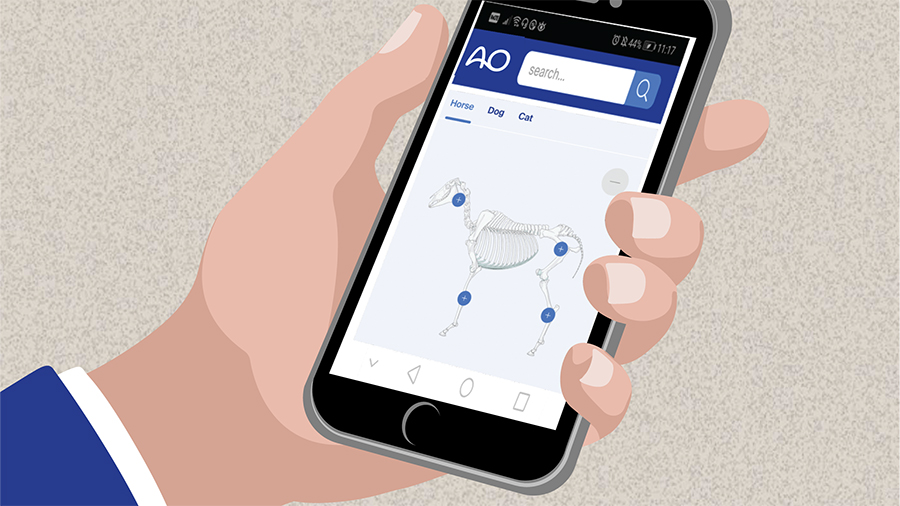
AO Surgery Reference
An internet-based resource for the management of equine, dog, and cat fractures.

FULL ACCESS MEMBERS ONLY
Journals and publications
A collection of publications and scientific journals hand picked for you.
FREE
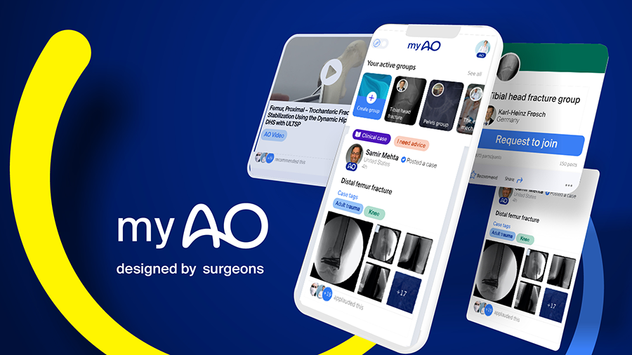
myAO
myAO is AO's digital network. You can connect, securely exchange knowledge with peers, and access leading clinical and scientific expertise.