
Managing chronic Monteggia fractures in dogs: Clinical insights for veterinary orthopedists

When I see a dog with chronic forelimb lameness, I know there’s a wide range of possible causes—but every so often, I’m reminded to look for the less obvious ones. Monteggia fractures, with their combination of ulnar fracture and radial head luxation, don’t show up often in practice. In this article, I’ll share my experience managing a large-breed dog with a chronic, bite-related Monteggia lesion, and walk through the practical steps that helped guide diagnosis, surgery, and recovery.
-
Read the quick summary:
- Dr Lisa Piras explains the diagnosis and surgical management of chronic Monteggia fractures in dogs, emphasizing the complexity of these cases.
- Timely diagnosis and adaptable surgical strategies, including ligament repair and temporary stabilization, are essential for good outcomes.
- Veterinary orthopedists benefit from recognizing subtle clinical and radiographic signs and employing advanced surgical and follow-up protocols.
- Ongoing discussion centers on sharing case experiences, surgical techniques, and best practices for postoperative care and rehabilitation.
Disclaimer: The article represents the opinion of individual authors exclusively and not necessarily the opinion of AO or its clinical specialties.
What is a canine Monteggia fracture?
A Monteggia fracture in dogs refers to a specific orthopedic injury pattern: a fracture of the ulna, usually in its proximal third, along with a luxation of the radial head at the elbow joint. While these injuries are less common than other forelimb fractures, their impact on elbow stability, limb function, and long-term mobility can be profound. In dogs, Monteggia lesions are often caused by high-energy trauma, such as vehicle accidents or falls, but can also result from bite wounds, an important consideration for any vet evaluating acute or chronic lameness with a history of trauma.
Failure to promptly identify and address both the ulnar fracture and the concurrent radial head luxation can lead to chronic pain, reduced range of motion, and progressive joint degeneration. Because the injury disrupts the congruence of the elbow, restoring anatomy is essential for optimal function.
Diagnosing chronic Monteggia fractures in dogs: key clinical and imaging clues
Accurate and timely diagnosis is the foundation for successful treatment. Dogs with chronic Monteggia fractures often present with severe, non-weightbearing lameness, marked pain upon elbow manipulation, and a restricted range of motion. In chronic cases, like the 12-year-old Giant Schnauzer referenced here, these signs may persist or worsen weeks after the initial injury, especially if the trauma was not recognized or properly managed (as with bite wounds).
Orthogonal and comparative radiographs are essential. Look for an ulnar diaphyseal fracture and displacement of the radial head, typically cranially or laterally. It’s easy to miss the luxation if you focus solely on the ulna, so a careful evaluation of elbow congruence is critical. Advanced chronicity leads to periarticular fibrosis and remodeling, which complicates both diagnosis and treatment. Awareness of this injury pattern can prompt early referral and improve prognosis.
Surgical solutions for chronic Monteggia fractures: when standard reduction isn’t enough
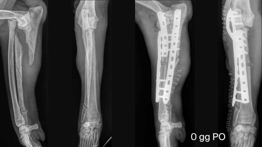
Managing chronic Monteggia fractures demands surgical flexibility. In chronic cases, anatomical reduction of the radial head is often blocked by fibrosis and altered bone surfaces. In our case, open reduction alone was insufficient, so a radial ostectomy was performed to shorten the radius and facilitate reduction.
Stabilization of the ulnar fracture was achieved with a locking compression plate (LCP), while the radial ostectomy was fixed with a cranial LCP. Intraoperative challenges are not uncommon—here, an iatrogenic rupture of the lateral collateral ligament required immediate attention. We restored elbow stability using two suture anchors (one on the humeral condyle and one on the radial head) connected with absorbable suture material. A contoured locking plate was also used for temporary trans-articular stabilization of the elbow.
These steps highlight the importance of adapting surgical techniques to the chronicity of the lesion and being prepared to address collateral ligament injuries and utilize temporary stabilization to protect the joint during early healing.
Maximizing recovery in challenging forelimb fractures
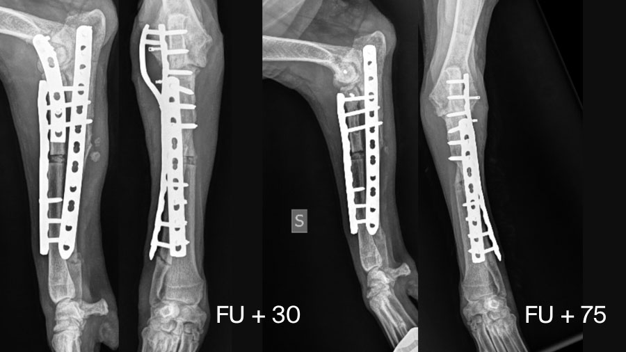
Postoperative management is just as critical as the surgery itself. Recovery is typically gradual. In this case, the dog showed steady improvement in limb loading and muscle mass over several weeks. Serial radiographs every two weeks monitored the healing of both the ulnar fracture and the radial ostectomy site. At 30 days, radiographic evidence supported removal of the trans-articular plate, which was performed around six weeks postoperatively.
Functionally, the patient regained good limb use, satisfactory elbow range of motion, and restoration of muscle mass. While physiotherapy is strongly recommended to enhance recovery, owner compliance may vary. Nevertheless, with vigilant monitoring and timely intervention, chronic Monteggia fractures can achieve positive outcomes.
What every vet should know about chronic Monteggia injuries
Here are some practical lessons I’ve learned from managing chronic Monteggia fractures that may help you in your own cases:
- Maintain a high index of suspicion for Monteggia fractures in any dog with forelimb lameness and a history of trauma, especially bites.
- Careful orthopedic and radiographic evaluation of the elbow joint is crucial—do not focus solely on the ulnar fracture.
- Chronic cases often require advanced surgical strategies such as radial osteotomy, ligament repair, and temporary trans-articular stabilization.
- Be prepared for intraoperative surprises and adjust your plan as needed; flexibility is key for optimal joint function.
- Close postoperative follow-up is essential, with regular radiographs and owner education about the importance of rehabilitation.
If you have managed similar cases or have questions about surgical techniques or postoperative protocols, I invite you to share your experience. Our profession advances most when we learn from each other’s challenges and solutions.
About the author:

Dr Lisa Piras graduated from the School of Veterinary Medicine at the University of Turin on April 26, 2005, with a thesis on “Use of the Ilizarov Technique for Fracture Treatment in Dogs and Cats,” supervised by Prof. Bruno Peirone. She obtained her professional license following the Board Examination at the University of Turin in November 2005. She later completed her PhD with a dissertation on “Limb Alignment,” under the supervision of Prof. Bruno Peirone and Prof. Derek Fox.
Currently, Dr Piras serves as an Assistant Professor at the University of Turin. She has been an invited speaker at numerous national and international conferences and courses, and in 2023, she became a Diplomate of the European College of Veterinary Surgeons.
You might also be interested in...
Small Animal Fracture Management
The aim of this Principles course is to introduce the principles of fracture management in small animals.

Lameness in Dogs online course
Develop and refine knowledge, skills, and attitudes in the diagnosis and decision-making of lameness in dogs.
Blog post: Beyond the Limp—diagnosis and treatment
Dr Stefan Scharvogel explains why a thoughtful, individualized approach is key to uncovering what lies behind the symptom.