
Beyond the limp: the art and science of diagnosing and treating canine lameness
BY DR STEFAN SCHARVOGEL
When a dog limps, it is never just a limp but rather a sign of something deeper. And although uncovering the root cause is not always straightforward, it is the critical first step toward effective treatment. A proper diagnosis requires a combination of keen observation, hands-on examination, and sometimes advanced diagnostics. It is not just about treating a symptom—it is about finding the underlying condition and choosing the best path forward. Here, Dr Stefan Scharvogel explains why a thoughtful, individualized approach is key to uncovering what lies behind the symptom.
Disclaimer: The article represents the opinion of individual authors exclusively and not necessarily the opinion of AO or its clinical specialties.
Before you can adequately address any medical condition, you need a diagnosis. This statement may seem like a truism, but diagnosis is the foundation of everything. Without it, you simply will not be able to establish and execute an effective treatment plan.
When it comes to lameness in dogs, the limping itself is not the diagnosis but merely a symptom—it is always the result of an underlying condition that causes it. For instance, a dog with a cruciate ligament rupture experiences pain in the affected leg. It will therefore attempt to put as little weight on the leg as possible, which results in limping. From a veterinarian’s perspective, once a dog like this presents at your practice, you need to ask yourself what steps you are to take in order to arrive at a diagnosis, and, in this specific example, to determine that the dog has a cruciate ligament tear.
Observe and touch
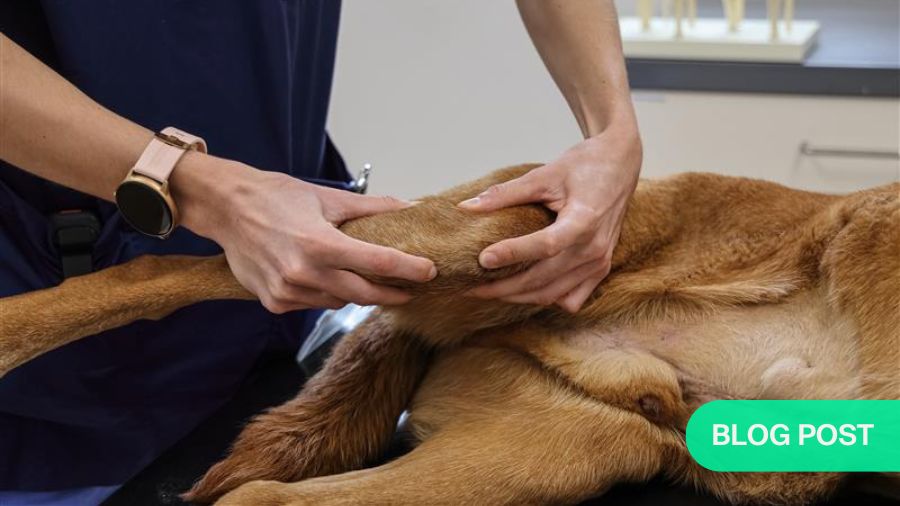
Indeed, the process itself is quite similar to what it would be with human patients. Unfortunately, many young veterinarians increasingly underestimate the importance of a thorough orthopedic examination. This means to truly look at the patient, for instance at its gait: how does the dog move? In some cases, you can establish quite a lot just from this. It also means to physically touch your patient. Physical contact can reveal so much. When you consult a doctor as a human patient, you may notice that hands-on examinations have become quite uncommon these days—and unfortunately, that is not always to the patient’s benefit. It is the same with animals.
My point is—and this is something that especially young veterinarians should take to heart—that we need to actually physically touch our patients if we are to conduct a comprehensive clinical examination. As a next step, we may need to undertake further diagnostics. This may involve imaging techniques like X-rays, CT scans, or MRIs. It might also entail a joint fluid analysis or blood tests. There is a wide array of options available.
Leveraging advanced diagnostics
One of the challenges is the fact that lameness in dogs can vary significantly in terms of severity. Sometimes, it will not be immediately obvious. You need to thoroughly assess the dog's different movements—standing, walking, trotting—with a trained eye in order to pick up on subtle signs. Another challenge of course is that the patient will not help you in any way. They cannot tell you where exactly they experience pain, and so you have to figure it out by yourself. Personally, I do not see this as a disadvantage, because what a patient tells you and what is actually going on are not always the same thing. What it does mean though is that you have to really engage with the issue.
But that should always be the case anyhow—you cannot not just rely on a simple complaint. For instance, when a human tells you that they have pain in their left knee, you should not immediately decide on an MRI and forego a thorough manual examination. Instead, you must first make sure that it truly is the left knee that is the source of the problem.
Only after a proper examination can you decide which additional tests are needed. After all, we do not want to perform procedures that are unnecessary, and unnecessarily costly. In some cases, I can take an X-ray for 50 euros and arrive at a diagnosis. Alternatively, I could go for an MRI, a magnetic resonance imaging scan, which costs around 1,400 euros, only to reach the same conclusion. And this is precisely what young veterinarians at the beginning of their careers need to internalize: for each patient, they should ask themselves what the right approach is. What is the best path forward for each individual patient?
Fortunately, recent technological advances have provided us with valuable new tools. One example is imaging: things like X-rays, CT scans, and MRIs, which have long been standard technologies in terms of human medicine, have also advanced significantly in veterinary medicine and are being applied regularly. Having said that, it is worth taking a global perspective and noting that there are still significant regional differences. In Europe or North America, obtaining an MRI is no major hurdle anymore—we do it whenever we believe it necessary. But if we look at regions like Africa, South America, or parts of Asia-Pacific, these technologies are not yet as readily available or commonly used.
Common causes of lameness
Although lameness in dogs has diverse underlying conditions, certain diagnoses are more common than others. For example, a cranial cruciate ligament rupture is extremely frequent. There are also some hereditary conditions that prevail in dogs. These are actively being addressed through breeding practices, but they can still lead to diseases that involve lameness. A prominent example is dysplasia, where, for instance, the joints in the elbow or the hip have developed abnormally. Elbow dysplasia and hip dysplasia are conditions we encounter relatively often. In other words, we have a number of hereditary conditions that we frequently need to identify and manage.
We also see trauma, of course. Dogs can suffer various types of injuries, for example during sports or just on a walk. They might twist a leg, and it does not always have to be a ligament tear or a fracture. These cases can sometimes be quite challenging to diagnose. So, there is a wide range of possibilities—quite similar, in fact, to what we see in humans.
The influence of size and lifestyle
An interesting aspect is the fact that there is some variation in terms of types of injury between different dog breeds, particularly between larger and smaller dogs. At the same time, there is a noticeable shift in the types of injuries we encounter, which has a lot to do with how and where people live and how they keep their dogs. For instance, with large dogs, we are seeing fewer fractures. In part, this is because cars are now built to be safer, reducing the risk of a broken leg if a dog collides with a vehicle. At the same time, dog ownership practices are changing: many dogs are no longer allowed to roam as freely as they used to, which again prevents certain types of injuries.
Then there are the so-called toy breeds. As urban areas around the world grow, dog sizes tend to shrink. In Japan, for example, people keep almost exclusively miniature dogs. In Europe, many people still live in rural areas, and especially in the southern countries, large dog breeds remain very popular. We are starting to see a trend toward smaller dogs here as well, but there are still significant differences.
Navigating the choices
When it comes to therapeutic approaches, a particular challenge lies in the decision between conservative and surgical treatment. Medicine is never a matter of 100—or zero—percent certainty. There are always differing opinions and multiple options. That is why the decision-making process is so critical: which direction should we take? With many conditions, we ask ourselves whether it really is strictly a surgical issue. A broken leg, for instance, will typically be treated surgically if we want it to heal fully—although we do sometimes see cases where a leg fracture will have healed reasonably well without surgery. But there are occasions where things are not so straightforward.
For instance, there are conditions where it is worth discussing whether a certain approach is truly beneficial, and where you are likely to encounter differing opinions. The diagnosis is just the first step. But even if a diagnosis has been firmly established, there often are various methods and approaches—all of them evidence-based—that can be applied . In many cases, the path forward therefore is not at all clear-cut.
“Trend” is an important term in this context, because treatment approaches often change over the years. These changes are not always evidence-based—they can sometimes be more like trends or fashions. That might not sound ideal, but it is a fascinating element of how practices evolve over time. Take elbow disorders, for example. Dogs frequently develop osteoarthritis of the elbow joint. However, whereas humans often develop arthritis due to wear and tear throughout their decades-long lifetime, this is unlikely in dogs given their much shorter lifespan of around 15 years. In veterinary medicine, we always assume that there is another underlying cause, for instance elbow dysplasia. And here, opinions diverge: some veterinarians argue for surgical intervention, while most papers and studies conclude that surgery is unnecessary and instead recommend conservative management with medication or physiotherapy. This also applies to other conditions: there are multiple treatment options, and each one is based on different considerations and perspectives.
Developments in surgical techniques
At the same time, new surgical methods are constantly in development. About 20 years ago, the introduction of arthroscopy marked a major breakthrough. In human medicine, computer-assisted surgical techniques are becoming increasingly common, but we have not yet reached that stage in veterinary medicine.
One significant challenge in terms of introducing new techniques, tools, and implants in veterinary practice is the vast size variation among our patients. All humans are generally similar in size, so a specific implant might be available in three or four sizes, with which you are able to cover nearly all patients. In contrast, our canine patients weigh between one and one hundred kilograms, presenting a much greater disparity. This makes it considerably more difficult to work with standardized solutions. However, a growing trend is the use of custom implants that are designed specifically for an individual patient’s needs and produced via 3D printing. In orthopedics, this is arguably the most important advancement of recent years.
Aftercare and rehabilitation
Success in the context of treating lameness in an animal ultimately means achieving the best possible outcome with a curative approach. Of course, there are cases where a complete cure is not possible. Joint arthritis is one example: unless you perform a joint replacement—which can be done in certain cases—this condition is not really curable. In such instances, we focus on achieving the best possible result. However, when a cure is attainable, that is always the goal—and in many cases, the full recovery of a patient is indeed possible.
This can be accomplished through either conservative or surgical treatment. Increasingly, however, aftercare and physical medicine also play a key role in veterinary medicine. Physiotherapeutic rehabilitation, or in some cases even physiotherapy as a standalone treatment, is standard practice these days.
Here, too, challenges arise due to the fact that we are treating animals, not humans. Our patients, of course, have their own will—and sometimes, they simply are uncooperative. For instance, hydrotherapy is a popular physiotherapeutic option: we let dogs walk in water, enabling them to exercise without bearing too much weight on their limbs. But if you have a dog that dislikes water, this particular approach is unlikely to work. You cannot persuade it or win it over with arguments, either. If a dog does not like water, it simply will not go in, and it will not move an inch.
Lifelong learning
If there was a piece of advice I could give to young colleagues in the field of veterinary medicine, it would be to get the best education that you possibly can. In my view, this is the key to truly understanding things, and it will continue to be so. This means to go through your formative years in the traditional sense and to take the time you need to get properly trained.
Because once you have gained that foundation, no one can take it away from you. I also think that it is equally crucial to never stop learning. Indeed, our professional code of conduct states that doctors must engage in continuous education—and I fully agree with that.
Ensuring the best possible medical care for our patients requires a commitment to lifelong learning. Never let up—your training is just the beginning. It is hard work, and it does not happen automatically. You will not have everything handed to you on a silver platter; that simply is not how it works. You need to have the drive and you must put in the effort yourself. Without that dedication, you will not achieve anything. That is how it always has been, and it is how it will always be.

AO VET Online Course—Lameness in Dogs: Diagnosis and Decision-Making
As a veterinarian looking to deepen your understanding and staying updated on the latest in diagnosing and treating lameness in dogs, consider enrolling in the AO Foundation’s online course on the issue. The six-week course offers comprehensive insights into the condition, including its causes, challenges in management, and advanced treatment options. Participants will benefit from expert knowledge, practical case studies, and interactive learning modules tailored to enhance their skills and knowledge in this critical area of veterinary medicine.
About the author:

Dr Stefan Scharvogel has had an extensive career in veterinary medicine and surgery. He conducted research at the Technical University of Munich (TU) and the University of Heidelberg. Following his general veterinary training at a private small animal clinic in Oldenburg, he earned his certification as a specialist veterinarian for small animals. He further advanced his expertise at the surgical department of the University of Leipzig’s Small Animal Clinic, where he became a certified specialist in veterinary surgery and achieved Diplomate status with the European College of Veterinary Surgeons (ECVS).
Scharvogel co-founded and was a member of the leadership team at the Tierklinik Haar. For 10 years, he served as a delegate for the Bavarian State Veterinary Chamber. He also chaired the German-speaking group of AO VET from 2012 to 2020 and is a founding member of the Veterinary Society of Surgical Oncology (VSSO). Additionally, he serves on the "Scientific Advisory Board" of Movora/Kyon and is an active member of numerous professional veterinary organizations.
You might also be interested in:
Lameness in dogs: diagnosis and decision-making
This 6-week online AO VET course focuses on enhancing decision-making skills specifically related to lameness in dogs and improving outcomes of canine surgery.

Discover more AO VET Courses and Events
Explore the upcoming courses, webinars or online events in your region or worldwide. You can download a list of all upcoming events in your region in PDF format.
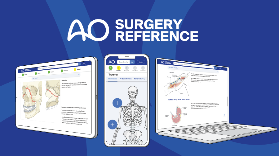
The AO Surgery Reference
The veterinary section contains content on fracture management in the horse, dog, and cat. Most anatomical regions are exclusively AO VET members only, some are open to all.
Orthopedic challenges in toy dog breeds
In the AO VET Blog, Lucas Beierer explains the importance of tailored treatment approaches in these uniquely rewarding pets due to their small size and bone structure.