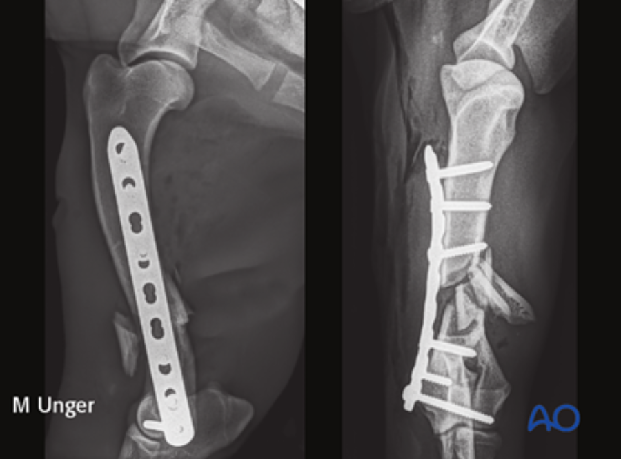
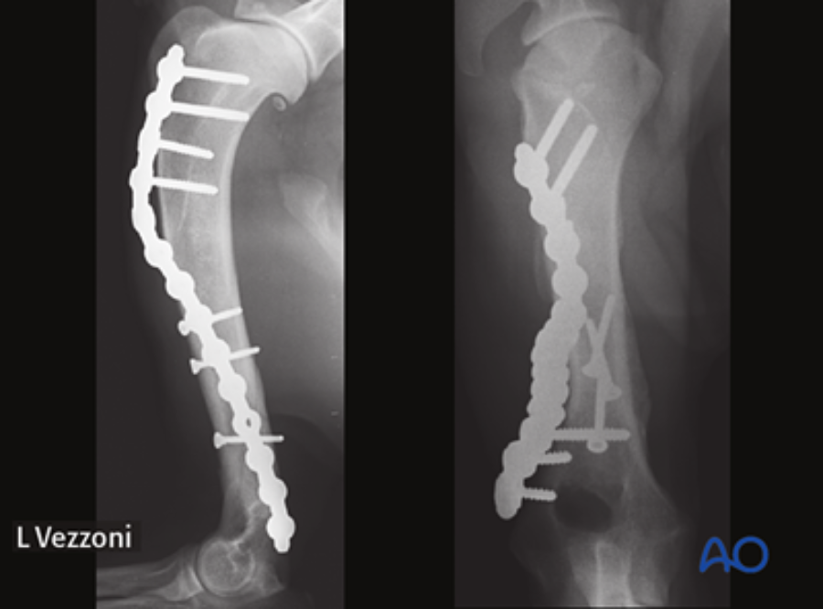
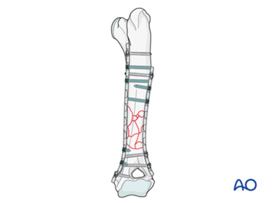
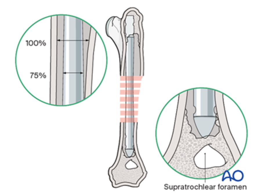
AO Surgery Reference's new dog humeral shaft fracture module
One-stop support for all levels of veterinary surgeons who treat dog humeral shaft fractures
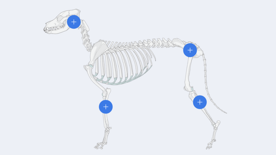
This recently released module on dog humeral shaft fractures offers a full range of reduction and fixation techniques, including a minimally-invasive one that preserves the maximum amount of periosteum while offering superior stability. While this module offers decision-making support for relative newcomers to veterinary trauma surgery, more experienced surgeons will also appreciate it as a very fast one-stop source of guidelines for diagnosis, indications, treatment and aftercare.
Dog humeral shaft module is exclusively available to AO VET members.
Not a member? Learn more about AO VET membership here.
What does the module include?
The module on this anatomical area was authored by Martin Unger (Germany). Matthew J Allen (UK) and Aldo Vezzoni (Italy) acted as editors.
This module includes sections on
- Fracture diagnosis
- Approaches to the humeral shaft
- Positioning
- Complications
- Wide selection of fracture fixation techniques tailored to specific fracture types
This module follows the AO VET classification for long bones. It offers an in-depth description of both lateral and medial approaches, along with a detailed exploration of a minimally invasive approach for the dog humeral shaft.
It encompasses both nonsurgical and surgical management techniques, providing detailed insights into the utilization of plates, intramedullary pins, and interlocking nails for fixation. Additionally, it offers supplementary reading materials that highlight the anatomical distinctions between dogs and cats, as well as the most prevalent complications that arise during humeral shaft fracture management.
What was the process of creating the module?
Review, analyze, incorporate, adapt, develop, illustrate and again, review!
Martin Unger: The module's development followed a meticulous, rigorous approach. Initially, following the fracture classification, we comprehensively reviewed the major textbooks, including the AO VET manual, to extract relevant fracture fixation methods. We then extensively analyzed our clinic's records and lectures from AO VET courses on humeral shaft fracture fixation. We incorporated other feasible fixation methods based on their potential value for specific fracture types.
More than 200 original illustrations
Detailed step-by-step illustrations include both cadaver approach photographs and standard images. To accelerate progress while ensuring a comprehensive and refined result, we collaborated with Carolina Salenius and Sabine Freiermuth through numerous video conferences and face-to-face meetings.
Once an initial version was completed, the module underwent a rigorous evaluation process. Esteemed professionals in the field, including Amy Kapatkin (executive editor), Aldo Vezzoni (general editor for thoracic limb), and Matthew Allen (AO VET’s incoming executive editor), meticulously reviewed it. Their invaluable insights and recommendations, including the addition of further case examples, were thoughtfully incorporated. This phase of review and quality control ensured a robust and comprehensive module even before the final version was produced.
The most interesting part of the new module?
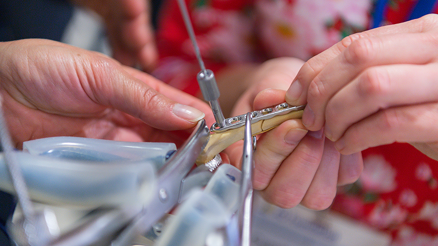
Matthew Allen: For me, the most interesting part of the module relates to the use of locking plates, especially in the context of minimally invasive surgical approaches. This method’s advantages include a combination of periosteal preservation and superior stability, which is achievable by locking the screw head directly into the plate. Where this approach is indicated, it is an excellent option for humeral shaft and humeral condylar fractures.
An old but still accurate adage
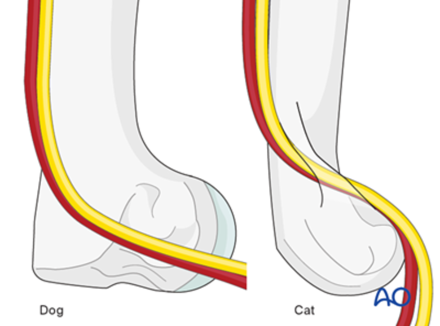
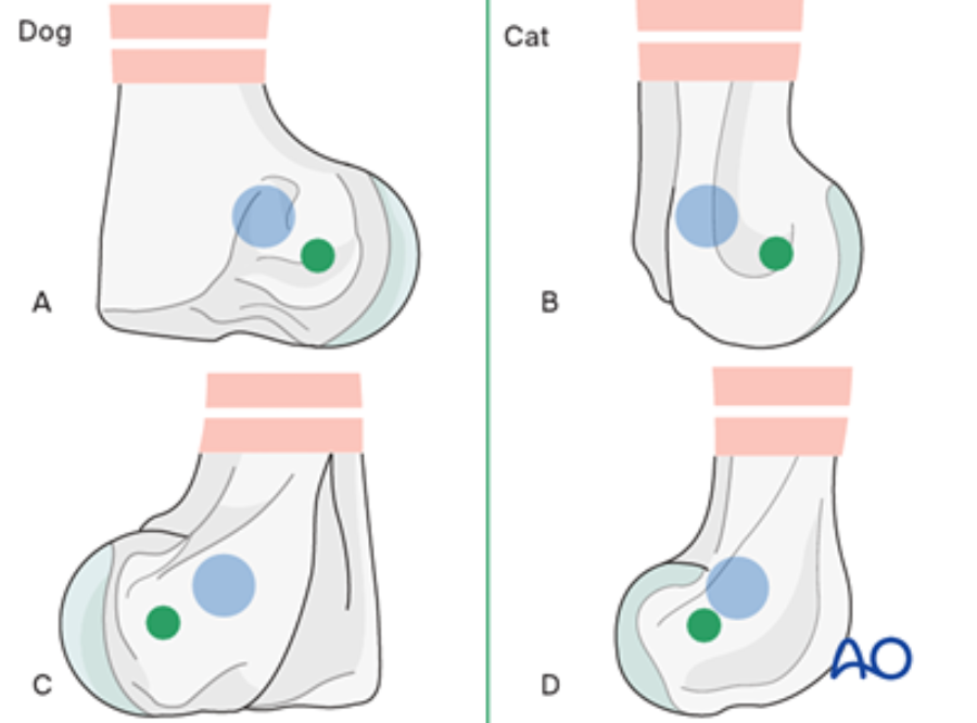
Cats are not small dogs. Differences in osseous anatomy and proportional bone geometry mean that, by treating cat humeral fractures as discrete entities, we can avoid complications such as pin penetration of the supracondylar foramen or joint violation by transcondylar screws.
The module highlights the anatomical differences between dogs and cats with illustrations.
Stay tuned!
The AO Surgery Reference team is working to complete surgical modules that cover the entire dog and cat skeletons. Modules are currently in preparation on the proximal radius and ulna, diaphyseal radius and ulna, distal radius and ulna, tarsus, and distal tibia. The distal humerus module will be published very soon!
You might also be interested in:
AO Surgery Reference
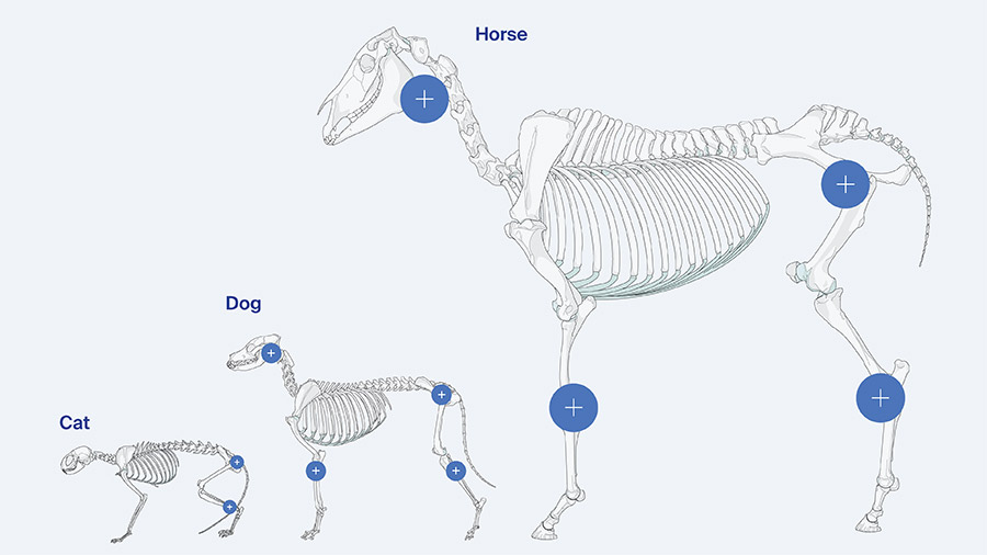
An internet-based resource for the management of equine, dog, and cat fractures.

