
Comprehensive patellar luxation in humans, horses, and dogs
BY DRS CHATCHANIN MAYURASAKORN, BARNY FRASER, AND CHALIKA WANGDEE

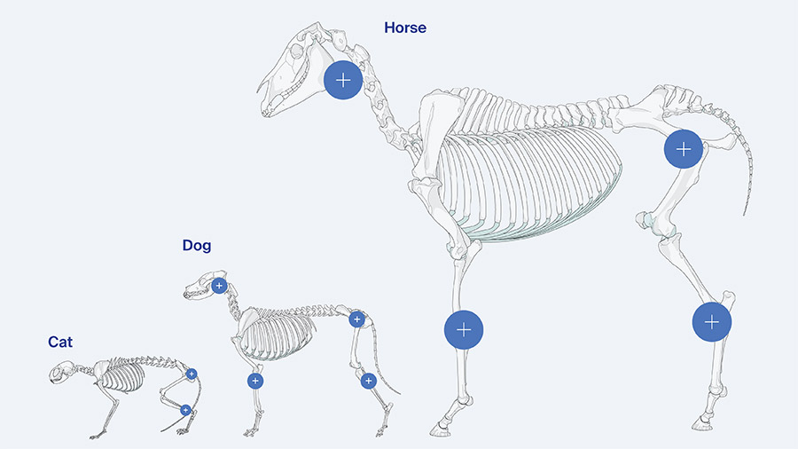
Patellar luxation and patellar instability are conditions encountered regularly in small animal practice, but they are far from unique to dogs. Similar disorders occur in humans and horses, and when viewed comparatively, they reveal a shared biomechanical foundation with species specific expressions. Across all species, the central problem is imbalance of the extensor mechanism, resulting in abnormal patellofemoral tracking. The way this imbalance manifests, progresses, and is treated depends heavily on anatomy, development, and functional demands.
Taking a comparative view has changed how I approach patellar luxation clinically. Rather than focusing on the patella alone, I now consistently treat the disease as a limb wide alignment problem. Human orthopedics, equine surgery, and veterinary small animal practice all arrive at the same conclusion through different paths: restoring extensor mechanism alignment is the cornerstone of successful treatment.
-
Read the quick summary:
- Surgeons discuss patellar luxation in humans, horses, and dogs, comparing anatomy, biomechanics, and clinical presentation across species.
- Shared extensor mechanism principles and the need for species specific strategies to prevent recurrent instability are key.
- Surgeons can apply comparative insights to improve case assessment, surgical planning, and long term outcomes in patellar instability.
- Ongoing discussion addresses how much correction is required beyond soft tissue repair when developmental or alignment factors are present.
Disclaimer: The article represents the opinion of individual authors exclusively and not necessarily the opinion of AO or its clinical specialties.
Shared biomechanical principles across species
Despite differences in limb orientation and locomotion, the patellofemoral joint functions as a pulley system in all species, optimizing the efficiency of the quadriceps mechanism. Patellar stability depends on the balance between osseous constraints, soft tissue restraints, and the direction of quadriceps pull. When these elements are misaligned, the patella no longer tracks centrally within the trochlear groove, leading to instability or frank luxation.
What differs is the direction of failure. In humans and horses, lateral luxation predominates. In dogs, and particularly small and miniature breeds, medial luxation is overwhelmingly more common. These patterns are not random; they reflect species specific limb alignment, muscle vectors, and developmental growth patterns rather than fundamentally different disease processes.
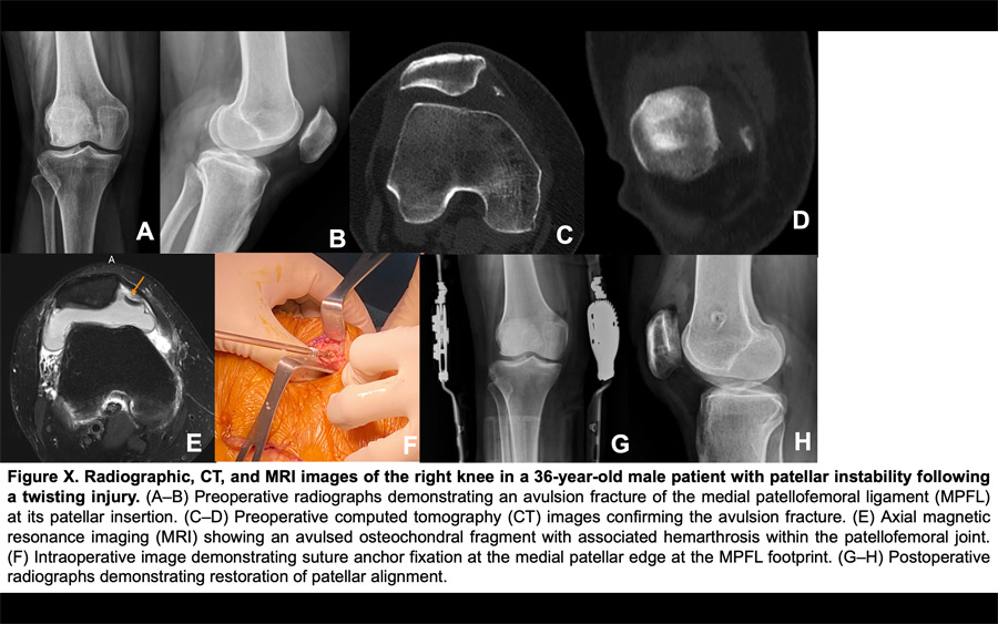
Patellar luxation in humans: lateral instability as the dominant pattern
In humans, patellar dislocation most commonly occurs laterally. This reflects the natural lateral orientation of the quadriceps pull relative to the mechanical axis of the limb. A combination of anatomical risk factors contributes to instability, including patella alta, trochlear dysplasia, increased Q angle, ligamentous laxity, and muscle imbalance, particularly insufficient function of the vastus medialis obliquus.
First time dislocations are frequently traumatic and often occur during twisting movements or direct impact with the knee flexed. Conservative treatment remains standard for initial events, consisting of reduction, bracing, restricted weight bearing, and structured physiotherapy.
Surgery is typically reserved for recurrent instability or for patients with clear anatomical predispositions that increase the risk of recurrence. Common procedures include medial patellofemoral ligament reconstruction, lateral release, tibial tuberosity medialization, and trochleoplasty when the groove is flat or convex.
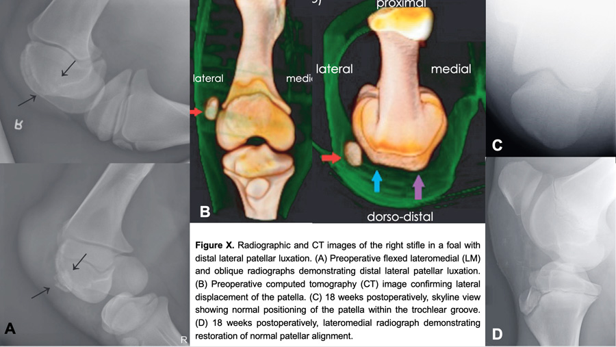
Patellar luxation in horses: rare but functionally significant
Patellar luxation in horses is uncommon, with only a small number of cases reported over decades. When it does occur, lateral luxation predominates and is most often associated with congenital maltracking, developmental abnormalities such as hypoplasia of the lateral trochlear ridge, or traumatic injury. Miniature horses and ponies appear overrepresented compared with full sized equids.
Surgical management in horses is constrained by size, biomechanics, and postoperative demands. Tibial tuberosity transposition, which is a mainstay in human and canine surgery, has been described only rarely in equids and is not routinely feasible. Instead, treatment typically focuses on soft tissue balancing through lateral release and medial imbrication, often combined with trochlear recession sulcoplasty when groove depth is inadequate. Prognosis varies, but outcomes are generally more favorable in foals and miniature horses than in adult athletic horses.
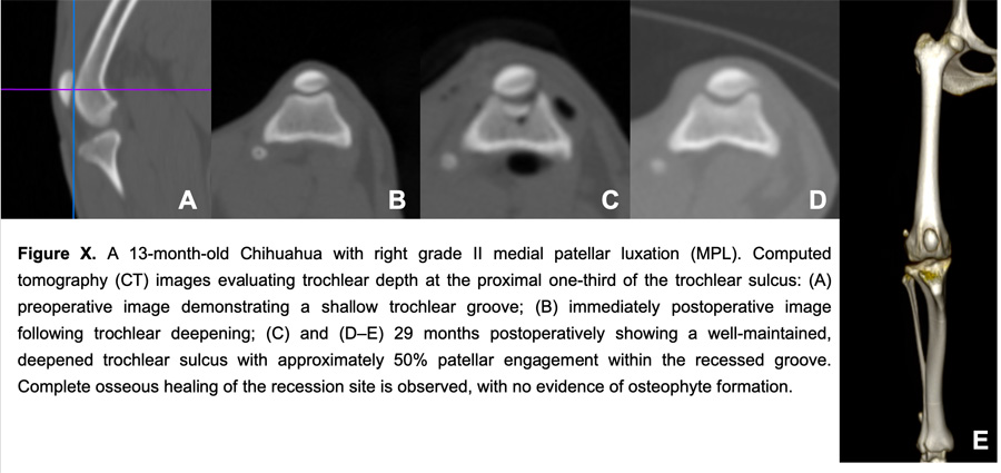
Patellar luxation in dogs: a developmental alignment disorder
In dogs, patellar luxation is predominantly medial and is best understood as a developmental orthopedic disorder rather than a traumatic one. The condition typically becomes apparent early in life and arises from malalignment of the quadriceps–patella–tibial axis. This malalignment alters limb biomechanics, leading to progressive dysfunction, intermittent or persistent lameness, and, in advanced cases, fixed luxation with marked femoral and tibial deformities.
Unlike humans, where traumatic events often initiate instability, most canine cases develop gradually as the animal grows. When luxation occurs in young dogs with open physes, delaying intervention allows angular and torsional deformities to worsen over time. What begins as a tracking problem can evolve into a complex multiplanar limb deformity that is far more challenging to correct later.
Treatment principles shared across species
Across humans, horses, and dogs, treatment goals are consistent:
- realign the extensor mechanism
- restore normal patellofemoral tracking
- stabilize the patella within the trochlear groove
- prevent recurrence
The challenge lies in selecting the appropriate combination of techniques for each species and each patient.
In humans, the emphasis is often on ligament reconstruction and selective bony procedures guided by well defined risk factors. In horses, soft tissue procedures dominate due to biomechanical constraints. In dogs, especially small breeds, surgery frequently requires a combination of trochlear deepening, soft tissue balancing, tibial tuberosity transposition, and corrective osteotomies to address underlying limb deformities rather than the luxation alone.
The role of alignment correction in canine cases
In my own practice, canine medial patellar luxation has reinforced the importance of comprehensive alignment analysis. Soft tissue procedures alone may be appropriate in very young patients to minimize physeal disruption, but in mature dogs, failure to correct femoral varus or tibial torsion often leads to recurrence. Corrective osteotomies are not optional add-ons; in fact, they are central to restoring normal biomechanics and durable patellar tracking.
This approach mirrors principles long established in human orthopedics, where isolated soft tissue reconstruction without addressing bony risk factors is associated with higher failure rates. Seeing the same pattern across species reinforces the value of alignment driven decision making.
Comparative insights for the practicing surgeon
Looking across species has sharpened my clinical judgment. Humans remind us to quantify risk factors and resist overtreatment after a single event. Horses illustrate the limits imposed by biomechanics and scale. Dogs demonstrate how developmental alignment errors can dominate disease progression. Together, they highlight that patellar luxation is never just a patellar problem.
For surgeons within the AO network, this comparative perspective supports a consistent principle: successful management depends on identifying and correcting the primary drivers of extensor mechanism imbalance. When we respect species specific anatomy while applying shared biomechanical principles, outcomes become more predictable and durable.
About the authors:

Dr Chatchanin Mayurasakorn, MD, FRCOST, is an Orthopaedic and Joint Replacement Surgeon in Thailand with special interests in orthopaedic traumatology and minimally invasive orthopaedic surgery. He completed his medical degree and orthopaedic training at Phramongkutklao College of Medicine, Thailand, and further pursued a Research Fellowship in Orthopaedic Traumatology at the University of Missouri-Columbia, USA.
Dr Chatchanin is actively involved in clinical practice, research, and academic activities, with publications in orthopaedic surgery and traumatology. His areas of interest include advanced fracture management. He is a member of the Thai Medical Council, the Thai Board of Orthopaedics, and the Royal College of Orthopaedic Surgeons of Thailand.
In addition to his clinical work, he has a strong interest in education and interdisciplinary collaboration, particularly in advancing musculoskeletal and joint surgery through innovation, research, and international cooperation.

Barny Fraser, BVSc MSc CertES(Orth) AFHEA DipECVS MRCVS, is an Associate Professor of Equine Surgery at Murdoch University in Western Australia and a Recognised Specialist since 2006 (ECVS, ABVS). He is primarily involved in clinical teaching of lameness and surgery in horses, with special interests in fracture repair and arthroscopic surgery. Research interests include the use of CT for pre-surgical diagnosis and planning as well as investigating longitudinal development of pre-fracture pathology using CT and MRI in young racehorses.
In addition to undergraduate, intern and resident training, his passion for clinical teaching led him to become initially AO VET Faculty and subsequently the Equine Representative on the Asia-Pacific Board of AO VET, where he hopes to help shape the future of post-graduate veterinary surgical education in the region.

Dr Chalika Wangdee is an Associate Professor of Veterinary Surgery at Chulalongkorn University, Thailand. She earned her DVM and MS from Chulalongkorn University and completed a PhD at Utrecht University, the Netherlands, focusing on patellar luxation. Her work centres on small animal orthopedic surgery, combining teaching, clinical service, and research.
Dr Wangdee was awarded an AO VET Fellowship in 2019, selected as a De Facto Diplomate of the Asian College of Veterinary Surgeons (AiCVS), and currently serves as President of the Thai Society of Veterinary Surgeons (TSVS) for 2023–2028. She heads the CU Innovation Center for Veterinary Clinical Training and practices in the Surgical Unit of the Small Animal Teaching Hospital.
She established the Soft Cadaver and Innovation Center (operational since 2020) and actively organizes advanced wet laboratory courses across Asia. Dr Wangdee joined the AO Faculty in 2022 and serves on the AO VET Asia Pacific Community Development Commission (2025–2028). She publishes widely and supervises graduate research in small animal orthopaedics.
You might also be interested in...
AO Surgery Reference
An internet-based resource for the management of equine, dog, and cat fractures.

AO VET Education
Delivering life-long education through courses and training worldwide.

Small Animal Fractures
Principles of fracture management in small animals: assessment, planning, and performance.
About AO VET
Advancing the practice of veterinary surgery to improve patient outcomes.