


Automated Deformity Analysis and Osteotomy Planning Tool in the Coronal Plane
Steffen Schröter, Julian Fürmetz, Jörg Harrer
The mediCAD AO Osteotomy software has been developed by the AO TC Deformity Correction Planning Task Force in collaboration with mediCAD® Hectec GmbH. It is the first fully automated 2D osteotomy planning tool based on long leg radiographs in the coronal plane. The software uses artificial intelligence to automatically detect relevant landmarks that are required for the calculation of all alignment parameters needed to analyze the deformity. According to the deformity, the software provides a recommendation whether single tibial, single femoral, or double-level osteotomy is preferable to correct the alignment. Furthermore, hinge point and cutting lines are automatically suggested and correction to a default target simulated.
Clinical problem
Knee joint preservation procedures have witnessed a resurgence of interest during the last 20 years. Congenital and posttraumatic deformities may lead to overloading of a compartment or even osteoarthritis. Osteotomies around the knee help to realign the limb, shift the load from the arthritic compartment to the intact compartment, and preserve the joint as an alternative to partial or total joint replacement.
Even in cartilage and meniscal procedures a physiological alignment is a prerequisite for success. Recent research also recommends performing osteotomies in ligament procedures to reduce the risk of revision.
In posttraumatic situations deformities may hinder an adequate bone healing because of biomechanical changes. Osteotomies can realign the limb, restore physiological biomechanics, and thus support bone healing.
The final limb alignment is a key predicting factor for favorable outcomes. Several steps are essential to achieve a perfect limb alignment. A significant contribution to the success of knee osteotomy is a thorough understanding of deformity analysis and surgical planning. There are two different steps: Deformity analysis enables the surgeon to identify the location of the deformity and choose the correct location and type of osteotomy, which is subsequently accurately planned.
Traditional deformity analysis and planning with hand drawings is time consuming, especially in cases in which more than one osteotomy is needed like double-level osteotomies. Since digital x-rays became the global standard, digital deformity analyzing, and planning are of substantial interest. Several digital planning software tools are available. A user-friendly interface is a vital feature of any software solution.
Development project
The mediCAD AO Osteotomy software has been developed by the AO TC Deformity Correction Planning Task Force in collaboration with mediCAD Hectec GmbH. It is the first fully automated 2D osteotomy planning tool based on long leg x-rays in the coronal plane. The software uses artificial intelligence to automatically detect relevant landmarks that are required for the calculation of all alignment parameters needed to analyze the deformity. According to the deformity, the software provides a recommendation whether single tibial, single femoral, or double-level osteotomy is preferrable to correct the alignment. Furthermore, hinge point and cutting lines are automatically suggested and correction to a default target simulated. The user can follow the workflow by accepting all recommendations unchanged, or edit them at any time, or even switch to a completely manual planning of the surgical procedure. Templating of plates and screws for fixation from various manufacturers is also possible. X-rays in several image formats can be used. The software makes it feasible to export the deformity, analyzing and planning with intermediate steps of the full procedure to be accessible in the operating room.
Availability of the mediCAD software solutions
Currently, the software must be installed on a client or clinical server to be available to the planning surgeons. A web-based version is expected by the end of 2022.
Details of planning
For digital planning of a deformity correction in the coronal plane, a long leg full weight-bearing x-ray in true AP is essential. A calibration ball or a scale is required to calibrate the planning and to measure wedge heights. A correct AP projection of the x-ray is the prerequisite to measure all angles accurately and must be controlled by the surgeon.
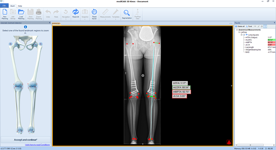
The software automatically detects all necessary landmarks (Fig 1). Based on their position (which can be adjusted manually) the software calculates all essential values (axis, joint lines, and angles) and delivers a detailed report. A traffic light system illustrates normal, intermediate, or pathological values.
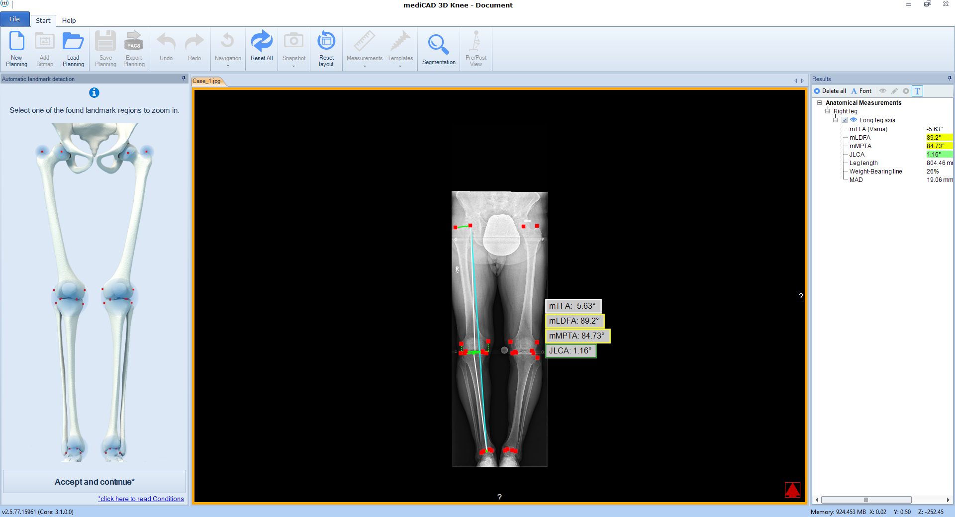 Fig 1 Detected landmarks and determined axes after automated segmentation.
 Following the deformity analysis, the software automatically recommends a surgical procedure, eg, medial opening wedge high tibia osteotomy, depending on the type of deformity (Fig 2). The user can follow the proposed procedure or choose individual options.
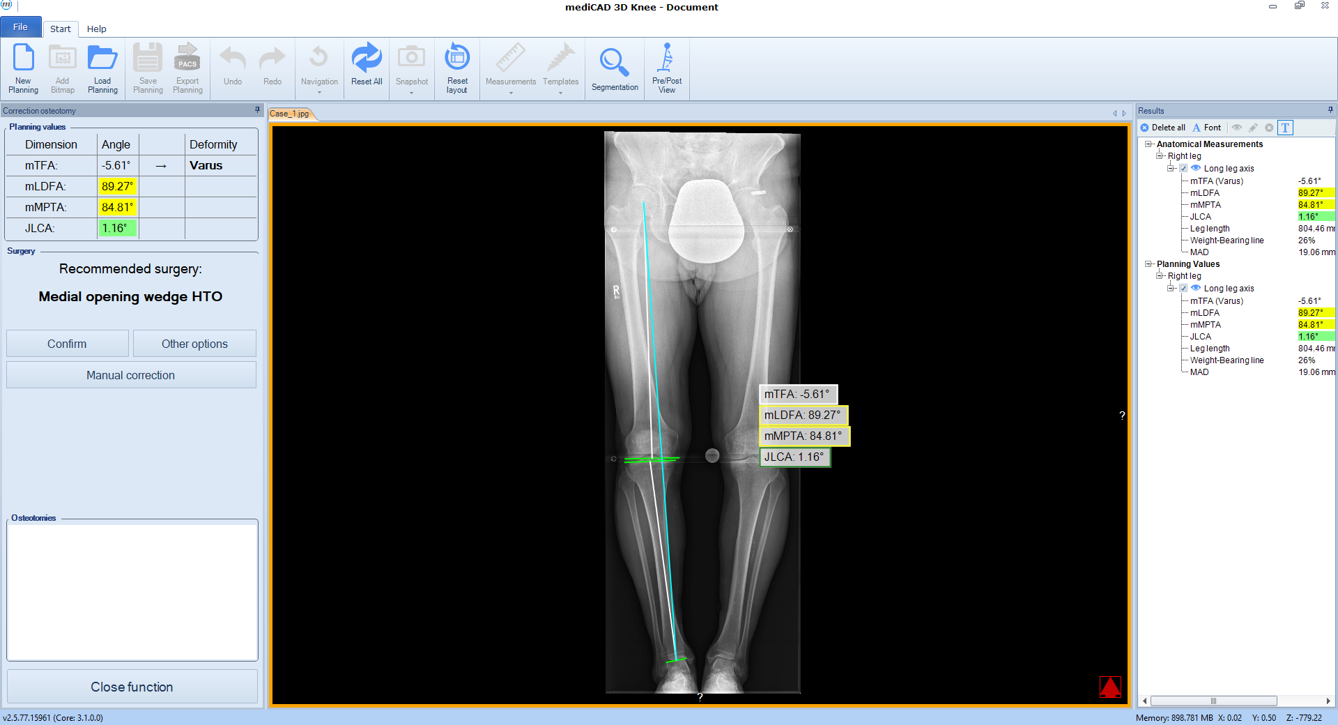 Fig 2 Deformity analysis with recommendation of osteotomy.
 Once the procedure is selected, the software automatically places the cutting line and hinge point for the chosen osteotomy adapted to the individual anatomical condition (Fig 3).
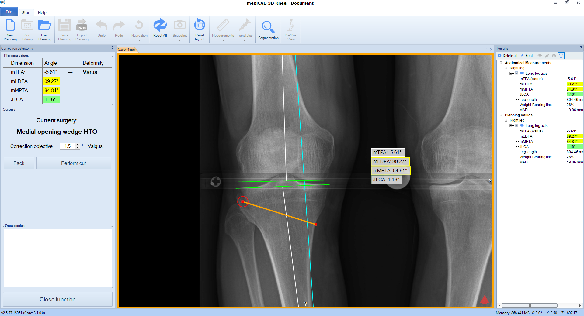 Fig 3 Proposed cutting line and hinge point.
 Next, deformity correction is visualized to the preferred new alignment. If a monofocal correction results in pathological joint angles, the implemented algorithm recommends double-level osteotomy and balance the two osteotomies between multiple constraints and normal values such as wedge heights, mechanical medial proximal tibial angle (mMPTA), mechanical lateral distal femoral angle (mLDFA), mechanical tibiofemoral angle (mTFA); (Fig 4).
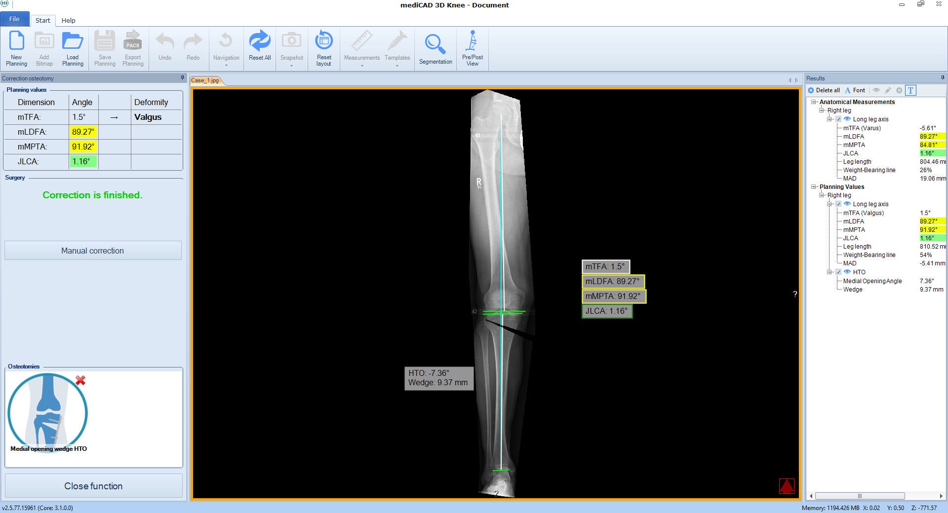 Fig 4 Simulation of proposed correction and resulting alignment.
 The Deformity Analysis and Osteotomy Planning Tools are offered to AO Trauma members at a discount of 20% for software licenses and 10% for services, such as on-site training, installation and alike, on the regular price as listed in official price lists in the country, where the AO Trauma member is located and working.
Please contact mediCAD if you are interested to learn more about the mediCAD osteotomy planning tool.
Case 1
(Case provided by Stefan Schröter)
A 33-year-old woman complained of pain in the lateral compartment after a normal working day. Sports activities were no longer possible. Free range of motion (ROM: 0/0/150°), ligaments in sagittal, and coronal planes were stable. There was no effusion. The patient had valgus deformity which was corrected with a distal femoral osteotomy.
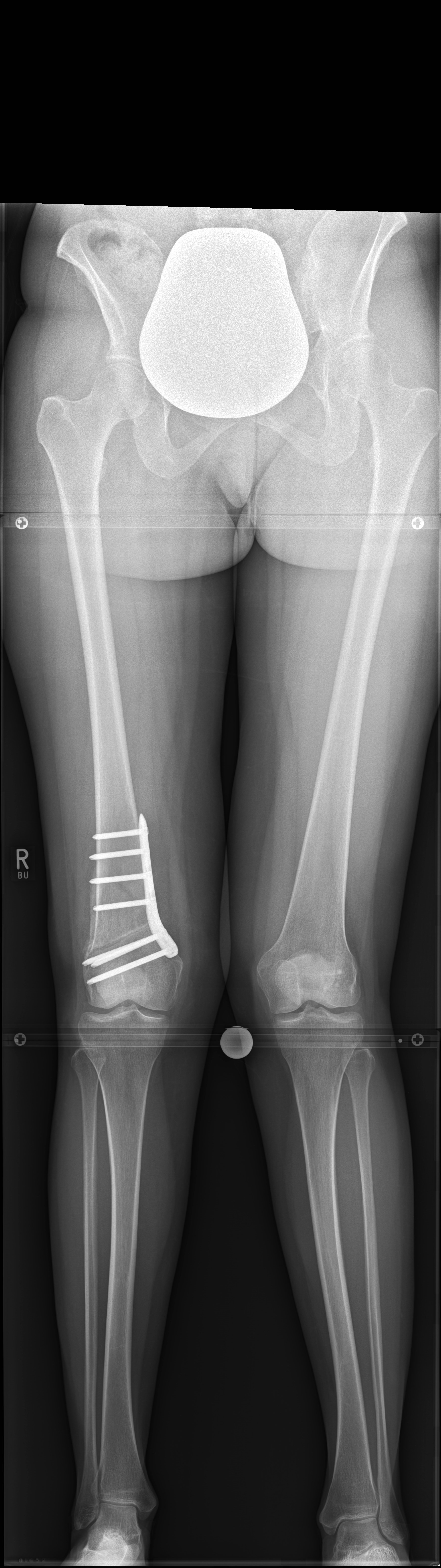 Fig 5: Pre-op radiograph.
 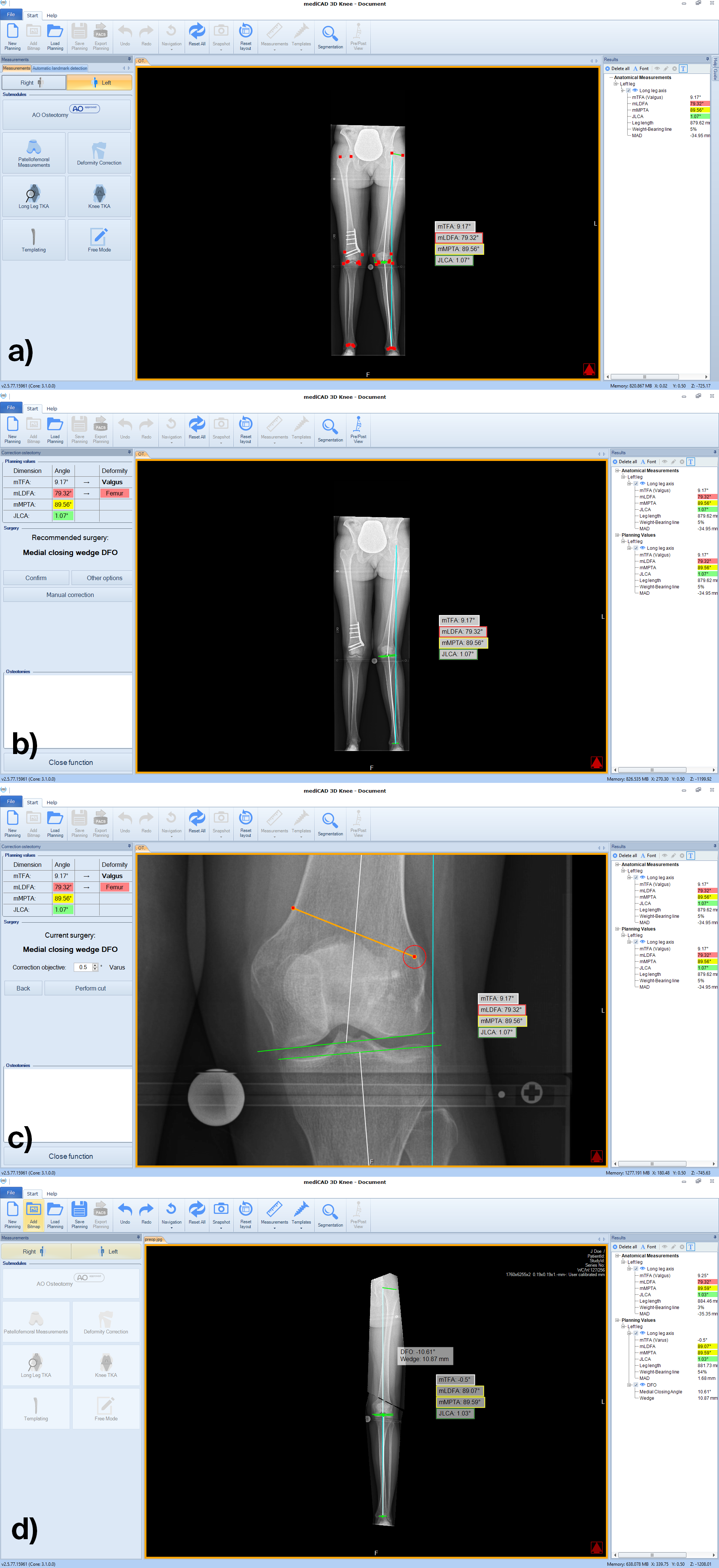 Fig 6a-d: Workflow based on pre-op radiograph.
 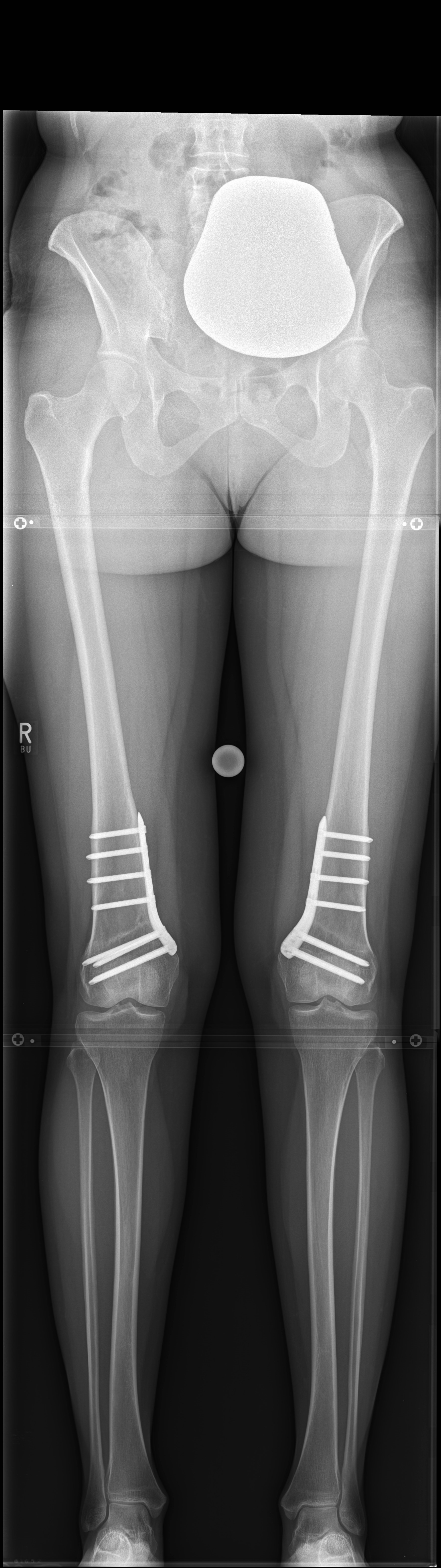 Fig 7: Post-op radiograph.
 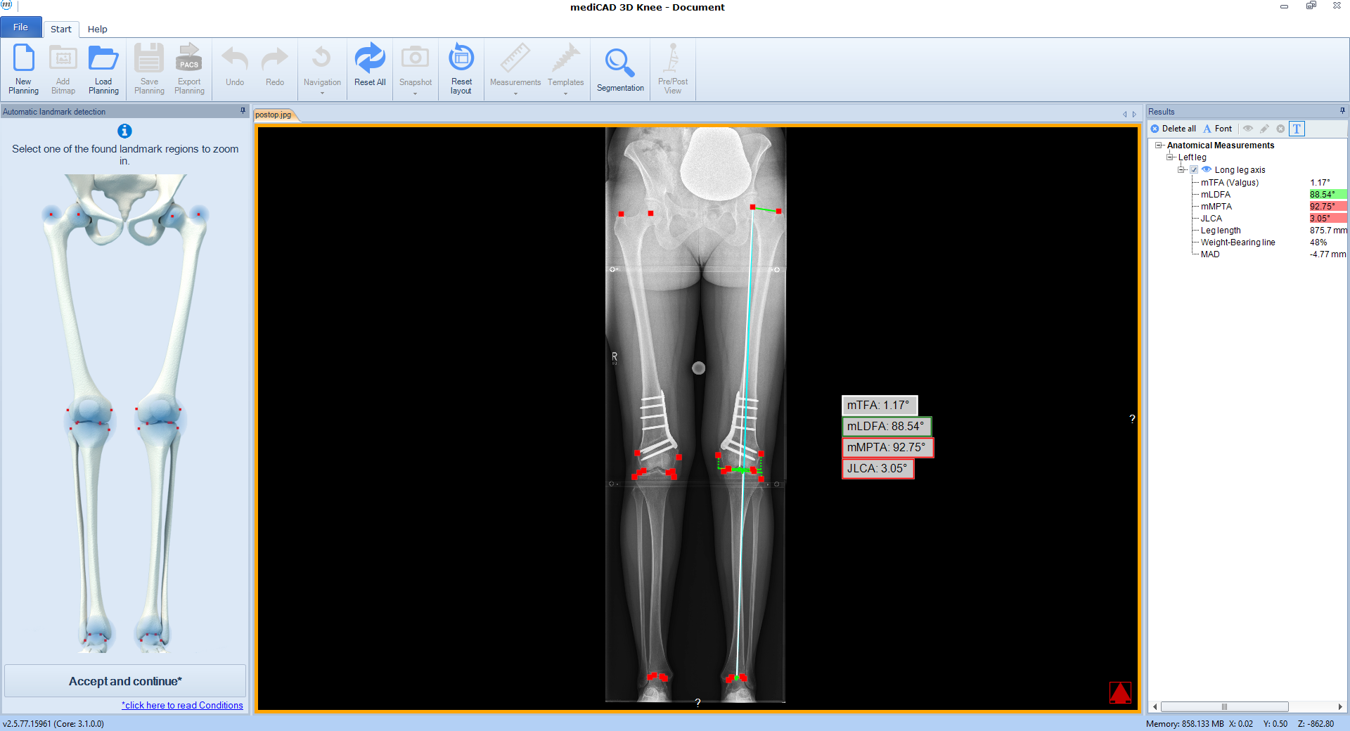 Fig 8: Post-op analysis in the software.
 Case 2
(Case provided by Steffen Schröter)
A 52-year-old man with a posttraumatic deformity 2 years after tibial plateau fracture AO/OTA 41C3.3e, PL, PM complained of pain after a normal working day. The patient was a heavy smoker. Activities like sports or hiking were discontinued. Surprisingly ligaments in the coronal and sagittal planes were stable. There was no effusion. Pain was localized in the medial compartment. The range of motion was limited to 0/0/130°. The deformity was corrected with a high tibial osteotomy.
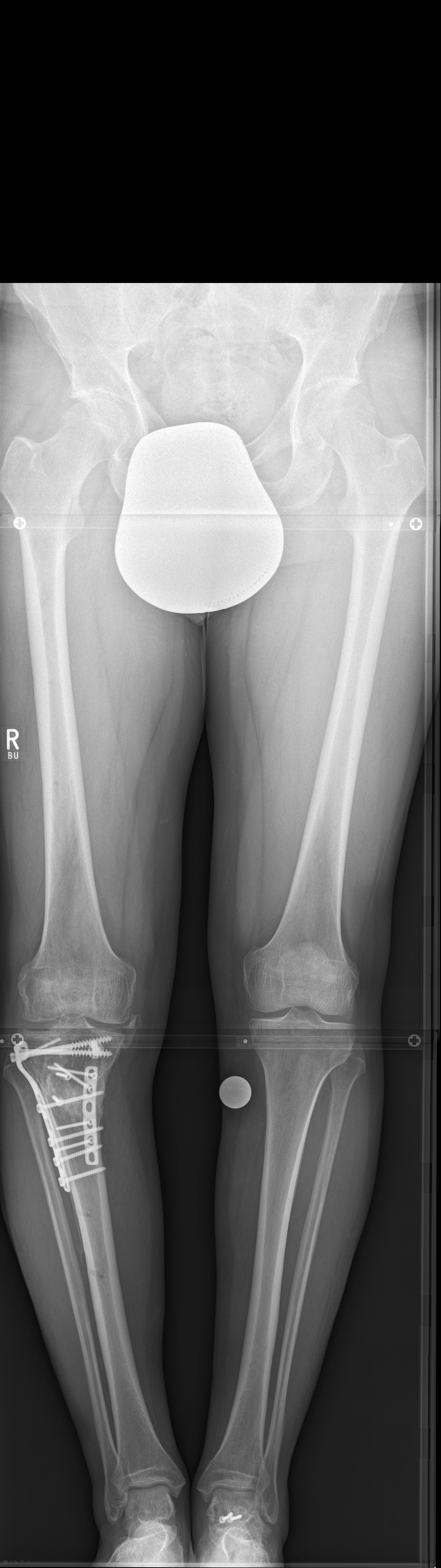 Fig 9: Pre-op radiograph.
 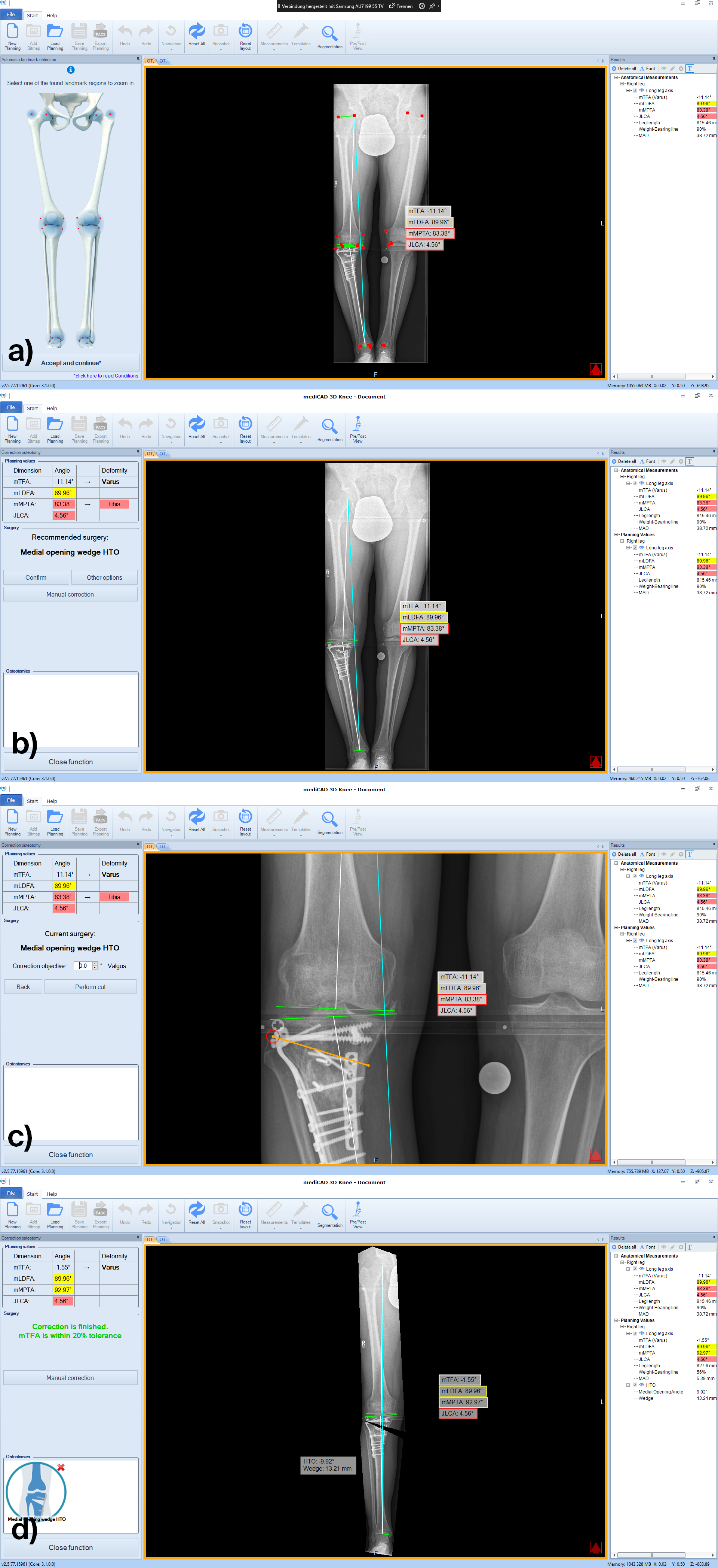 Fig 10a-d: Workflow based on pre-op radiograph.
 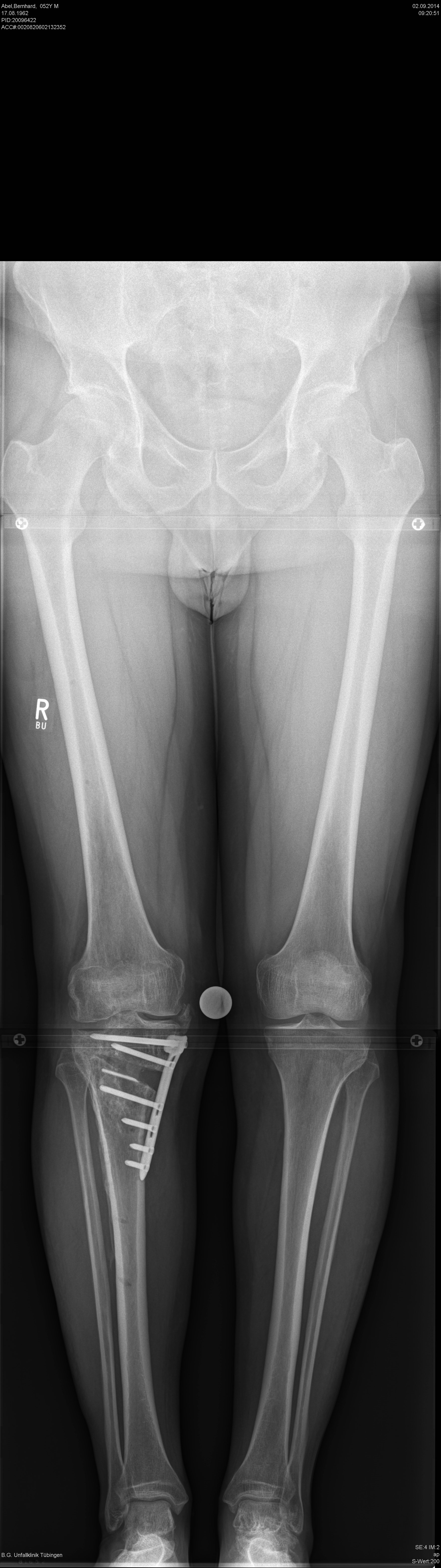 Fig 11: Post-op radiograph.
 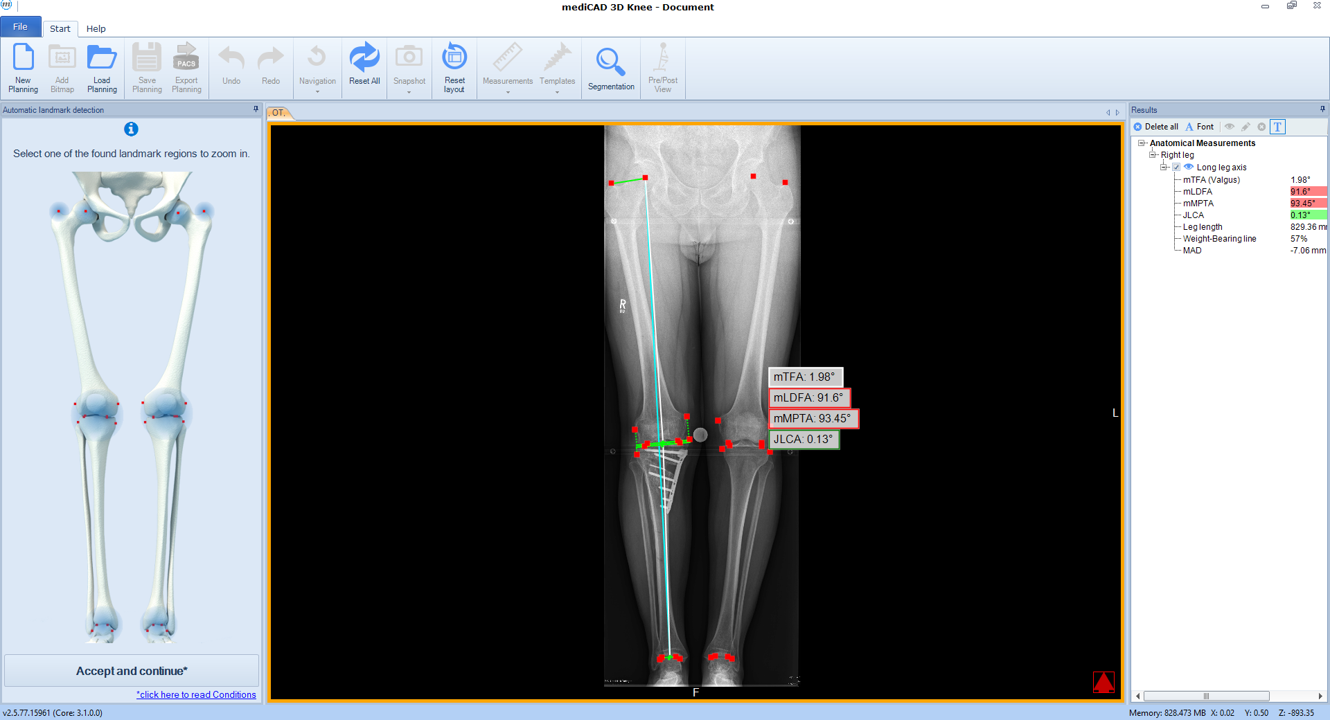 Fig 12: Post-op analysis in the software.
 Case 3
(Case provided by Steffen Schröter)
A 52-year-old man, a heavy worker was still able to work, however he was administered pain killers. During weekends only brief bike rides were tolerable. He reported an effusion and on that evening further daily activities were not possible. He complained of pain in the medial compartment. Excessive bowing legs were noticeable. Ligaments were stable in the sagittal plane but there was a medial instability in the coronal plane because of damage to the cartilage. He underwent double-level osteotomy for correction of the leg alignment.
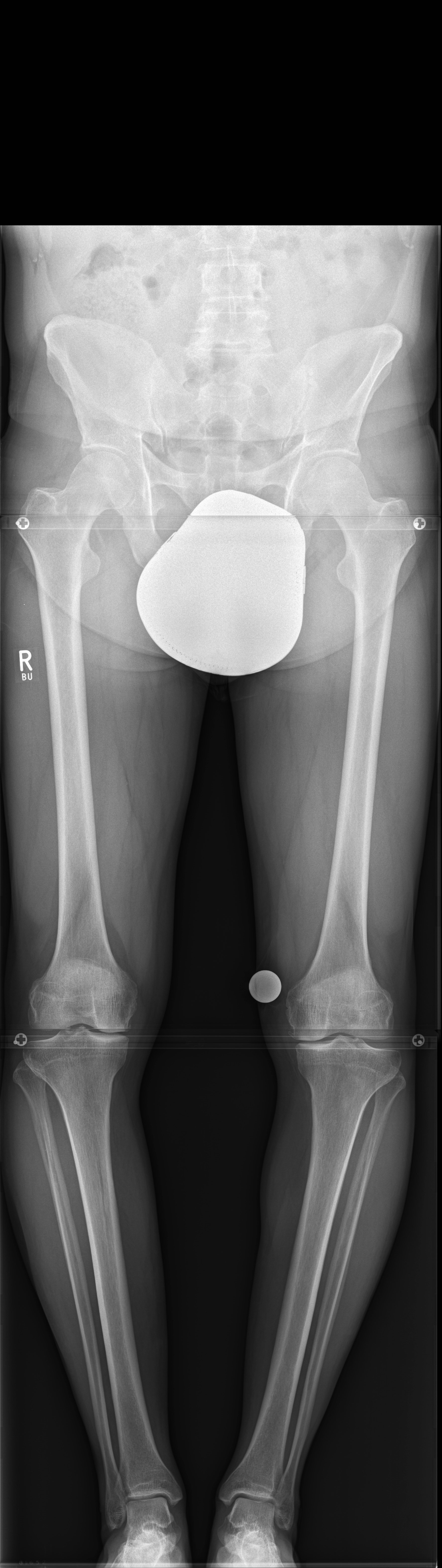 Fig 13: Pre-op radiograph.
 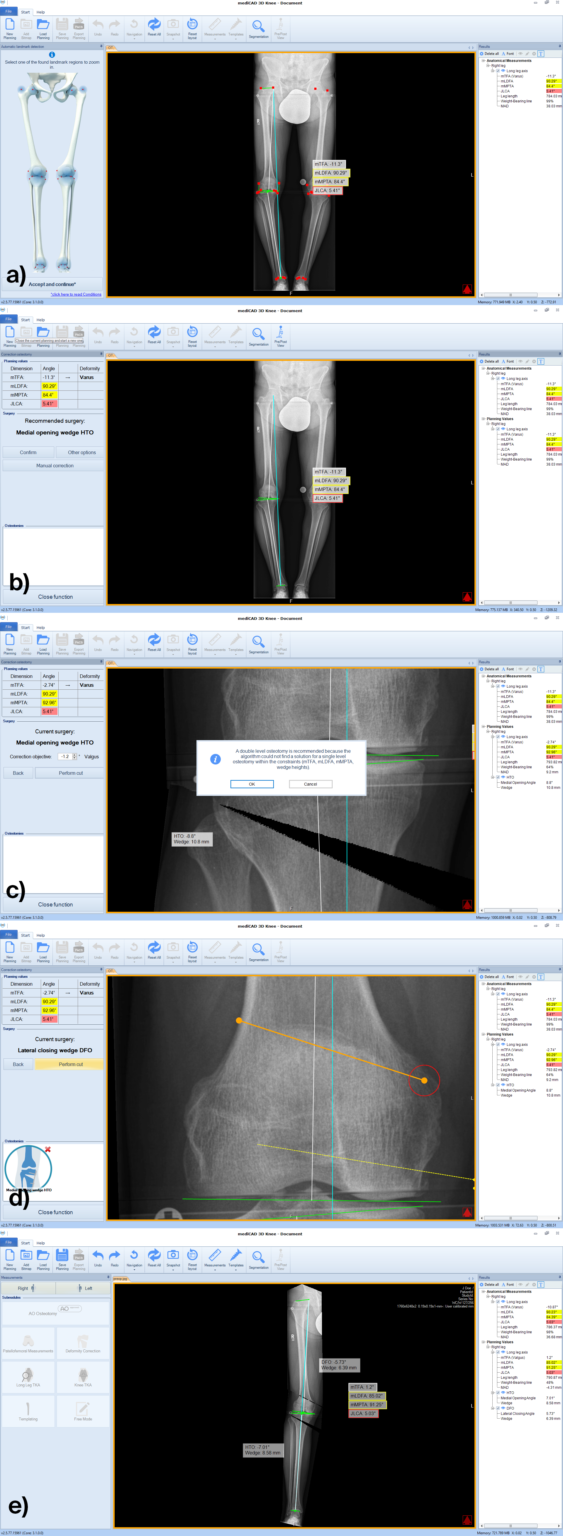 Fig 14a-d: Planning workflow with recommendation to change to double level osteotomy.
 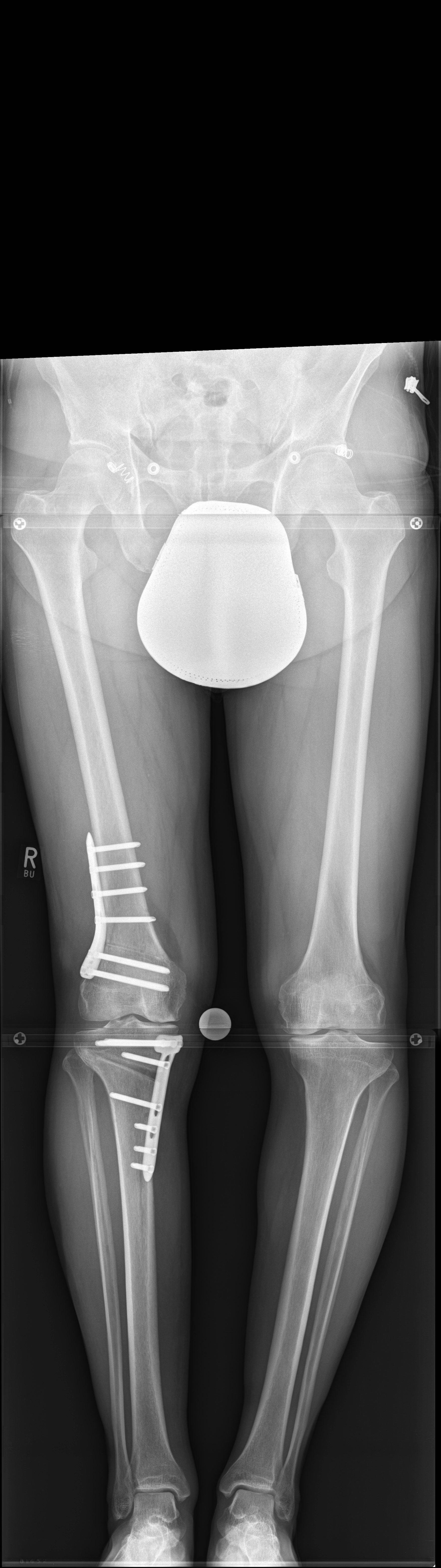 Fig 15: Post-op radiograph.
 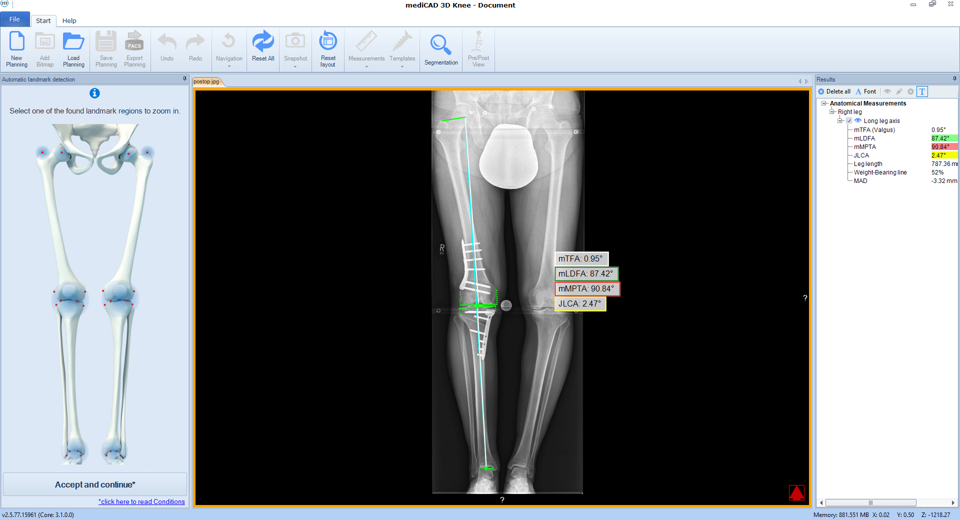 Fig 16: Post-op analysis in the software.
 Showcasing a distal femur osteotomy case
Showcasing a high tibial osteotomy case
Showcasing a double-level osteotomy case
Hazards and labeling
Due to varying countries’ legal and regulatory approval requirements, consult the appropriate local product labeling for approved intended use of the products described on this website. All devices on this website are approved by the AO Technical Commission. For logistical reasons, these devices may not be available in all countries worldwide at the date of publication.
Legal restrictions
This work was produced by AO Foundation, Switzerland. All rights reserved by AO Foundation. This publication, including all parts thereof, is legally protected by copyright.
Any use, exploitation or commercialization outside the narrow limits set forth by copyright legislation and the restrictions on use laid out below, without the publisher‘s consent, is illegal and liable to prosecution. This applies in particular to photostat reproduction, copying, scanning or duplication of any kind, translation, preparation of microfilms, electronic data processing, and storage such as making this publication available on Intranet or Internet.
Some of the products, names, instruments, treatments, logos, designs, etc referred to in this publication are also protected by patents, trademarks or by other intellectual property protection laws (eg, “AO” and the AO logo are subject to trademark applications/registrations) even though specific reference to this fact is not always made in the text. Therefore, the appearance of a name, instrument, etc without designation as proprietary is not to be construed as a representation by the publisher that it is in the public domain.
Restrictions on use: The rightful owner of an authorized copy of this work may use it for educational and research purposes only. Single images or illustrations may be copied for research or educational purposes only. The images or illustrations may not be altered in any way and need to carry the following statement of origin “Copyright by AO Foundation, Switzerland”.
Check www.aofoundation.org/disclaimer for more information.
If you have any comments or questions on the articles or the new devices, please do not hesitate to contact us.
“approved by AO Technical Commission” and “approved by AO”
The brands and labels “approved by AO Technical Commission” and “approved by AO”, particularly "AO" and the AO logo, are AO Foundation's intellectual property and subject to trademark applications and registrations, respectively. The use of these brands and labels is regulated by licensing agreements between AO Foundation and the producers of innovation products obliged to use such labels to declare the products as AO Technical Commission or AO Foundation approved solutions. Any unauthorized or inadequate use of these trademarks may be subject to legal action.

AO ITC Innovations Magazine
Find all issues of the AO ITC Innovations Magazine for download here.


Innovation Awards
Recognizing outstanding achievements in development and fostering excellence in surgical innovation.