VET LCP Notched Head T-Plate
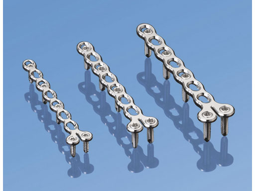
The VET LCP notched head T-plates are indicated for metaphyseal fractures of long bones in toy through to small-sized dog breeds, and cats. Its primary usefulness, however, is for the commonly observed distal radial and ulnar fractures in toy and miniature dog breeds. These fractures typically occur only 12 cm from the distal end of the bone. These fractures in the toy and miniature dog breeds require rigid fixation as an experimental study suggested that a decreased vascular supply to the distal radius in small breeds compared to other breeds may contribute to an increased incidence of delayed and nonunions [1].
Because of the fracture location resulting in a small distal bone fragment, there is only sufficient room to place two screws with a T-plate design. The notch in the T-plate allows independent contouring of the plate at this level, facilitating its placement in the area of the radial groove adjacent to the articulation (for the extensor carpi radialis muscle tendon). The ability to place two locked screws at this level also enables a more secure fixation. Finally, the combination hole of this LCP allows standard screw fixation to be applied, where the fracture site can be compressed by loading the screw in the dynamic compression unit portion of the hole, thus increasing the fracture stability. Secondly, angulation of a screw also may be performed in the dynamic compression unit hole so as to avoid the fracture line. Locking screws subsequently may be applied in the threaded portion of the remaining combination holes, as indicated.
Reference
1 Welch JA, Boudrieau RJ, Djardin LM, et al (1997) The intraosseous blood supply of the canine radius: implications for healing of distal fractures in small dogs. Vet Surg; 26(1):5761.
These plates have exactly the same design as the LCP condylar plates for use in humans. They are available in three sizes, 2.0, 2.4, and 2.7 mm. All three sizes feature seven combination holes in the shaft and two locking holes in the plate head. The plates may be cut in order to obtain the desired length. The low profile of the plates with rounded edges and a highly polished surface minimizes soft-tissue irritation (for example, under the extensor tendons of the distal radius). The plates can easily be bent or cut with the respective instrumentation.
Yorkshire Terrier
(Case provided by Randy J Boudrieau, North Grafton, USA)
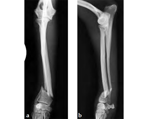
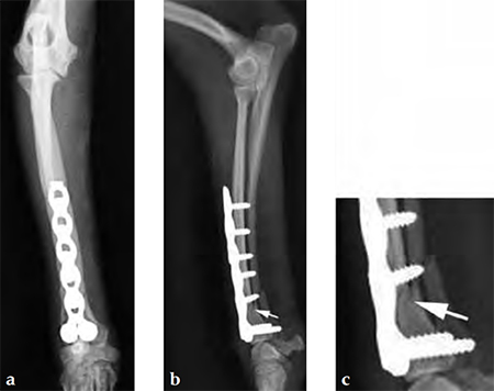
A 1-year-old female Yorkshire Terrier (1.7 kg) fractured the right distal radius and ulna (similar to a Colles fracture in humans). Open reduction and internal fixation was performed using a 7-hole 2.0 mm VET LCP notched head T-plate (a 7-hole plate was cut to eliminate the proximal two combination holes). The fracture was reduced and the distal bone fragment secured with two locking screws; the notch in the T-plate allowed independent contouring of this portion of the plate adjacent to the articulation. Compression was then applied across the fracture by loading one screw (second screw-hole proximal to the fracture); a second standard screw was then secured adjacent to the fracture (angled away/proximal to the fracture). The remaining screws were placed in a locked fashion. Despite the attempt at anatomical reduction, a slight (~1 mm) step was observed in the craniocaudal reduction (Fig 3). No fixation was applied to the ulna. A soft, padded bandage was applied to the forelimb (distal to the elbow joint) for minimal support and to control swelling.
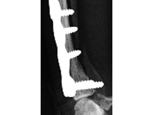
The dog was discharged from the hospital 2 days postoperatively with instructions for strict exercise restriction; the dog was ambulatory on all four limbs at this time, although lame on the right forelimb. The bandage was to be removed in about 57 days. The dog did very well and was reevaluated 8 weeks postoperatively, at which time radiographs revealed a healed radius and atrophic ulna (Fig 4 - the ulnar atrophy is the norm in the canine for this repair in these small dog breeds). The dog was fully ambulatory and without lameness at this time; however, there was a slight decrease in full flexion at the antebrachiocarpal jointagain, this is the norm with this repair due to the surgical dissection and plate placement under the extensor tendons, which does not cause any functional deficit in the dog. The dog was gradually returned to full activity over the ensuing 2 weeks. It is recommended that these dogs not be allowed to jump from heights (eg, bed, chair) as they are at increased risk for this fracture, regarding the opposite limb in this patient.
Small Bones, Small Plates: Clinical Application of Mini LCP
Hazards and labeling
Due to varying countries’ legal and regulatory approval requirements, consult the appropriate local product labeling for approved intended use of the products described on this website. All devices on this website are approved by the AO Technical Commission. For logistical reasons, these devices may not be available in all countries worldwide at the date of publication.
Legal restrictions
This work was produced by AO Foundation, Switzerland. All rights reserved by AO Foundation. This publication, including all parts thereof, is legally protected by copyright.
Any use, exploitation or commercialization outside the narrow limits set forth by copyright legislation and the restrictions on use laid out below, without the publisher‘s consent, is illegal and liable to prosecution. This applies in particular to photostat reproduction, copying, scanning or duplication of any kind, translation, preparation of microfilms, electronic data processing, and storage such as making this publication available on Intranet or Internet.
Some of the products, names, instruments, treatments, logos, designs, etc referred to in this publication are also protected by patents, trademarks or by other intellectual property protection laws (eg, “AO” and the AO logo are subject to trademark applications/registrations) even though specific reference to this fact is not always made in the text. Therefore, the appearance of a name, instrument, etc without designation as proprietary is not to be construed as a representation by the publisher that it is in the public domain.
Restrictions on use: The rightful owner of an authorized copy of this work may use it for educational and research purposes only. Single images or illustrations may be copied for research or educational purposes only. The images or illustrations may not be altered in any way and need to carry the following statement of origin “Copyright by AO Foundation, Switzerland”.
Check www.aofoundation.org/disclaimer for more information.
If you have any comments or questions on the articles or the new devices, please do not hesitate to contact us.
“approved by AO Technical Commission” and “approved by AO”
The brands and labels “approved by AO Technical Commission” and “approved by AO”, particularly "AO" and the AO logo, are AO Foundation's intellectual property and subject to trademark applications and registrations, respectively. The use of these brands and labels is regulated by licensing agreements between AO Foundation and the producers of innovation products obliged to use such labels to declare the products as AO Technical Commission or AO Foundation approved solutions. Any unauthorized or inadequate use of these trademarks may be subject to legal action.
AO ITC Innovations Magazine
Find all issues of the AO ITC Innovations Magazine for download here.
Innovation Awards
Recognizing outstanding achievements in development and fostering excellence in surgical innovation.


