Sterile Artificial Skull
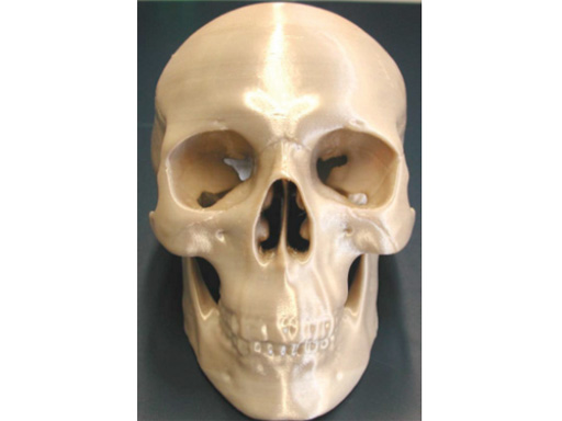
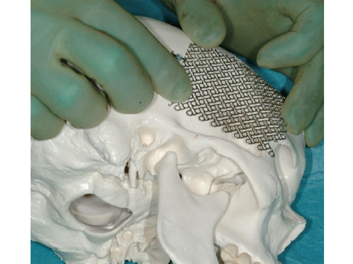
The new artificial sterilizable skull is the first autoclavable, one-piece human skull model intended for aiding initial plate contouring in the operating room, and for visualizing facial structures.
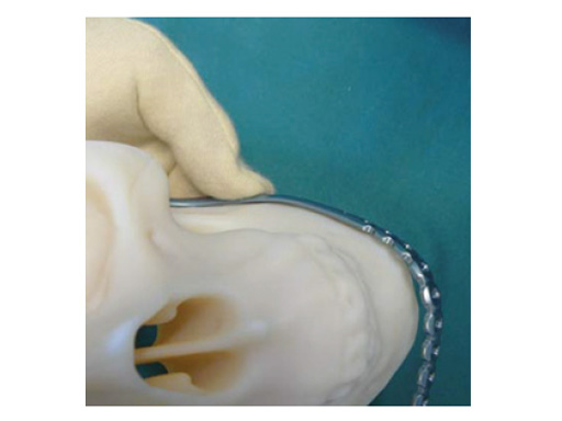
There is a need for sterile artificial skulls for any craniomaxillofacial (CMF) surgeon who provides CMF reconstructions. The clinical reality is that surgeons worldwide have individual methods to address this need due to the difficulty and lack of anatomical reference points in cases with large bone defects. However, the skull model needs to be mechanically strong for intraoperative contouring of plates, meshes, and biomaterials.
The patients anatomy is essential for a proper and adequate reconstruction of the defects. Improper contouring of the plates may lead to complications resulting in functional and/or aesthetic problems that require secondary surgery.
This sterile artificial skull model is an approximation of the average craniofacial anatomy, originating from data from up to 2000 analyzed CT scans. These scans were used to develop the matrix midface preformed orbital plates and the matrix mandible preformed reconstruction plates (Fig 2), launched in 2008 and 2009, respectively. The manufacturing material is polyphenylsulphone, which allows the skull to be steam sterilizable, reusable, cleanable, and biocompatible.
This new productthe sterile artificial skullcontributes to a more appropriate hard-tissue reconstruction in the CMF region, especially in cases of trauma, tumor, and craniofacial deformity. It provides a unique aid, always available and ready to use even in emergency cases.
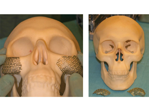
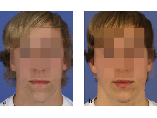
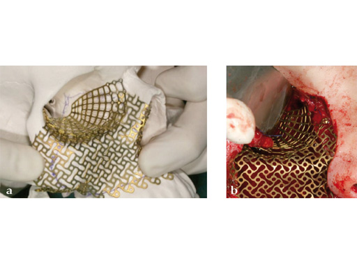
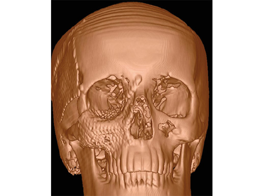
Case 1: Camouflaging of deficient zygomatic prominences by mesh augmentation. a 19-year-old man with a typical-angle class iii appearance is shown preoperatively (fig 4a) and postoperatively (fig 4b). after preoperative planning, a bimaxillary osteotomy with advancement of the upper jaw and backwards movement of the lower jaw following bisagittal split osteotomy was performed. alternative options to augment the malar bone are surgical osteotomy of the malar bone with outwards movement of the zygomatic prominences, high le fort i osteotomy with onepiece movement of the maxilla together with the zygomatic prominence, using other biomaterials than meshes to augment these prominences. in this case, the average prominence in an adult caucasian patient was mimicked by contouring a 0.6 mm thick, 3-D mesh using the artificial skull.
Case provided by Nils-Claudius Gellrich, Hannover, Germany
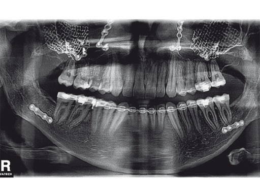
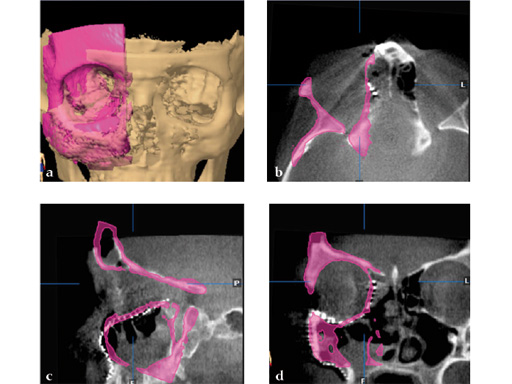
Case 2: A 19-year-old woman had an extended sinunasal carcinoma in the right maxillary sinus area. fig 2 shows four views of the virtual preplanned computer-aided design; the reconstruction is represented in pink (fig 2a). The three remaining multiplanar views show the overlapping images of the virtual preplanned reconstruction (pink) and reconstruction result performed by using two individualized 3-D meshes (one for a three-wall-reconstruction of the orbit and one for reconstructing the right midfacial prominences).
Cases provided by Nils-Claudius Gellrich, Hannover, Germany
Hazards and labeling
Due to varying countries’ legal and regulatory approval requirements, consult the appropriate local product labeling for approved intended use of the products described on this website. All devices on this website are approved by the AO Technical Commission. For logistical reasons, these devices may not be available in all countries worldwide at the date of publication.
Legal restrictions
This work was produced by AO Foundation, Switzerland. All rights reserved by AO Foundation. This publication, including all parts thereof, is legally protected by copyright.
Any use, exploitation or commercialization outside the narrow limits set forth by copyright legislation and the restrictions on use laid out below, without the publisher‘s consent, is illegal and liable to prosecution. This applies in particular to photostat reproduction, copying, scanning or duplication of any kind, translation, preparation of microfilms, electronic data processing, and storage such as making this publication available on Intranet or Internet.
Some of the products, names, instruments, treatments, logos, designs, etc referred to in this publication are also protected by patents, trademarks or by other intellectual property protection laws (eg, “AO” and the AO logo are subject to trademark applications/registrations) even though specific reference to this fact is not always made in the text. Therefore, the appearance of a name, instrument, etc without designation as proprietary is not to be construed as a representation by the publisher that it is in the public domain.
Restrictions on use: The rightful owner of an authorized copy of this work may use it for educational and research purposes only. Single images or illustrations may be copied for research or educational purposes only. The images or illustrations may not be altered in any way and need to carry the following statement of origin “Copyright by AO Foundation, Switzerland”.
Check www.aofoundation.org/disclaimer for more information.
If you have any comments or questions on the articles or the new devices, please do not hesitate to contact us.
“approved by AO Technical Commission” and “approved by AO”
The brands and labels “approved by AO Technical Commission” and “approved by AO”, particularly "AO" and the AO logo, are AO Foundation's intellectual property and subject to trademark applications and registrations, respectively. The use of these brands and labels is regulated by licensing agreements between AO Foundation and the producers of innovation products obliged to use such labels to declare the products as AO Technical Commission or AO Foundation approved solutions. Any unauthorized or inadequate use of these trademarks may be subject to legal action.
AO ITC Innovations Magazine
Find all issues of the AO ITC Innovations Magazine for download here.
Innovation Awards
Recognizing outstanding achievements in development and fostering excellence in surgical innovation.


