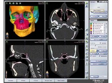
iPlan CMF 3.0
Computer technology has been utilized in CMF surgery for some time. Besides the improved ways of accurate positioning of implants during the operation (navigation), two additional aspects in computer-assisted surgery (CAS) are of great importance: First, new technologies allow for a precise preoperative planning procedure; second, CAS serves as an unrivaled quality control instrument postoperatively with great teaching quality.
The planning software iPlan CMF 3.0 allows for extensive CMF planning and bridges the gap between preoperative planning and intraoperative navigation. Streamlined, time-saving functionalities and automated processes have been added to prior versions aiding the surgeon in the two most important aspects of his procedures: accuracy and time.
Among a variety of features are:
- Virtual simulation of the surgical treatment
- Automatic segmentation of structures based on an anatomical atlas
- Transfer of complete virtual plan into the OR without loss of data for intraoperative realization
- Import of preformed orbita reconstruction plates via STL
- Generation models for customized implants
Preparation: easy alignment of DICOM datasets
The first step in using iPlan CMF 3.0 is the upload of the patients DICOM data. However, before the actual planning begins, the dataset needs to be aligned correctly. The midsagittal and the Frankfort horizontal plane serve as reference points and mirroring planes. The alignment can be done with a drag and drop movement of the dataset with computer mouse. As an additional feature it is possible to display two sides of the patient in one view, allowing for comparison of the anatomical structures.
Time saving atlas-based automatic segmentation
Following the correct alignment the next required step in the procedure is the creation of reconstruction templates for the areas of interest. In current solutions this was a complex and as a result time consuming process for the surgeon requiring his precise outlining with numerous mouse clicks. With its new automatic segmentation feature, iPlan CMF 3.0 segments objects accurately within minutes. This feature is based on an anatomical atlas and allows for simultaneous selection of several objects at once.
Finally the Smart Shaper feature enables the surgeon to perform further manual manipulation of the objects in 3-D until the dataset fully matches meets his requirements.
In addition to the enhanced efficiency in the planning process due to the significant time saved, the internal atlas also prevents pseudo-foramina which may happen to appear in areas with thin boney structures, ie, the orbit.
Mirroring of sides in unilateral defects
iPlan CMF 3.0 offers another feature which allows for easy mirroring of segmented objects along a defined plane. This is particularly versatile in cases with unilateral defects where the surgeon can use the anatomical structures of the nonaffected side as a template to plan the reconstruction of the injured side: By a click of the mouse segmented structures can easily be mirrored along the mid-sagittal plane and overlay the defect giving clear landmarks in distances, relationships, and projections. The surgeon is then able to again manipulate and position the segments by hand until they align giving a precise prediction for the reconstruction. If older image data of the patient is available this would even be possible in cases where both sides of the facial bone structure are damaged.
Export and import of STL data for CMF planning
Naturally the surgeons requirements for a planning tool do not end with a precise display of the anatomical structure itself, rather, it is of utmost importance to see if and how the chosen implants will help to achieve the desired reconstruction. To that end another important feature of this software tool is the ability to import STL data. This feature allows to import implants such as the preformed orbital plates (see "Preformed orbital plates" on page 11) into the patients planning dataset. This aids the surgeon in checking the correct implant size that is required for the defect as well as defining its optimal position simply by moving it in the dataset on the screen.
At the same time, STL files can also be exported after the planning procedure is finished, ie, to utilize the data for rapid prototyping or the production of patient-specific implants eliminating the need to produce physical models beforehand.
The final operation plan can be transitioned easily into the navigation system without switching software applications, thus avoiding potential software incompatibilities and lost data. Lastly iPlan 3.0 can be used as an objective quality control instrument helping the surgeon to compare the postoperative result with the original plan by use of best-in-class
image fusion technology.
CAS Planning in CMF Cases
Hazards and labeling
Due to varying countries’ legal and regulatory approval requirements, consult the appropriate local product labeling for approved intended use of the products described on this website. All devices on this website are approved by the AO Technical Commission. For logistical reasons, these devices may not be available in all countries worldwide at the date of publication.
Legal restrictions
This work was produced by AO Foundation, Switzerland. All rights reserved by AO Foundation. This publication, including all parts thereof, is legally protected by copyright.
Any use, exploitation or commercialization outside the narrow limits set forth by copyright legislation and the restrictions on use laid out below, without the publisher‘s consent, is illegal and liable to prosecution. This applies in particular to photostat reproduction, copying, scanning or duplication of any kind, translation, preparation of microfilms, electronic data processing, and storage such as making this publication available on Intranet or Internet.
Some of the products, names, instruments, treatments, logos, designs, etc referred to in this publication are also protected by patents, trademarks or by other intellectual property protection laws (eg, “AO” and the AO logo are subject to trademark applications/registrations) even though specific reference to this fact is not always made in the text. Therefore, the appearance of a name, instrument, etc without designation as proprietary is not to be construed as a representation by the publisher that it is in the public domain.
Restrictions on use: The rightful owner of an authorized copy of this work may use it for educational and research purposes only. Single images or illustrations may be copied for research or educational purposes only. The images or illustrations may not be altered in any way and need to carry the following statement of origin “Copyright by AO Foundation, Switzerland”.
Check www.aofoundation.org/disclaimer for more information.
If you have any comments or questions on the articles or the new devices, please do not hesitate to contact us.
“approved by AO Technical Commission” and “approved by AO”
The brands and labels “approved by AO Technical Commission” and “approved by AO”, particularly "AO" and the AO logo, are AO Foundation's intellectual property and subject to trademark applications and registrations, respectively. The use of these brands and labels is regulated by licensing agreements between AO Foundation and the producers of innovation products obliged to use such labels to declare the products as AO Technical Commission or AO Foundation approved solutions. Any unauthorized or inadequate use of these trademarks may be subject to legal action.
AO ITC Innovations Magazine
Find all issues of the AO ITC Innovations Magazine for download here.
Innovation Awards
Recognizing outstanding achievements in development and fostering excellence in surgical innovation.


