Curvilinear Distractor 2.0 mm
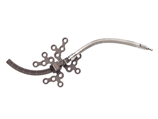
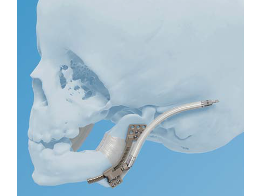
The new curvilinear distraction system is an internal distraction osteogenesis device that gradually advances the mandible along a curved trajectory of distraction. It addresses the clinical need for an internal mandible distractor that lengthens the mandible in both vertical and
horizontal planes. It can be speculated that simultaneous expansion of the mandible in two vectors also results in a significantly increased retroglossal airway and therefore is of particular benefit in very severe cases of mandibular hypoplasia [1].
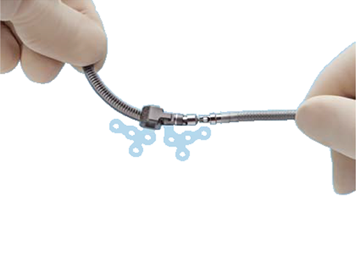
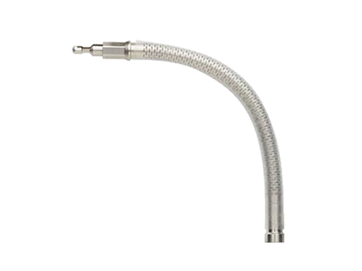
The system features various curved distractors which are fixed to the mandible with either locking or nonlocking 2.0 mm bone screws. All the distractors accept removable, flexible, or rigid extension arms from the CMF distraction system (see TK News 2/08) which move the point of activation to a location that is easily accessible with the activation instrument. The extension arms can be removed during the consolidation phase without a surgical procedure.
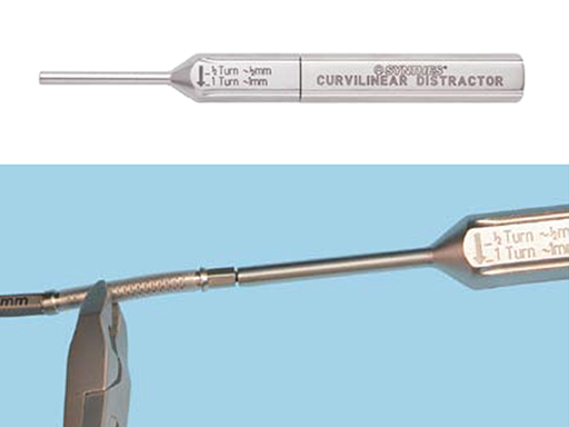
The distractor track is 35 mm long and can be cut to the desired length by the surgeon to accommodate individual patient needs. The underside of the track is etched to indicate the location where to cut to achieve the desired length. The end of the track is crimped to create a functional stop to prevent the distractor assembly from separating when fully distracted.
The curvilinear distractor is offered in left and right assembles in several different radii of curvature, including 30, 40, 50, 70, and 100 mm. The distraction radius corresponds to the distance in millimeters from the center of a circle to the outer circumference of the circle. A smaller radius results in a tighter curvature of distraction as opposed to a larger radius which is closer to a straight path of distraction. The distractor is also offered in a straight version.
Curvilinear distractor bending templates are available for preoperative planning. They can be used with anatomical bone models to perform model surgery. The bending templates translate along the curved track.
They are available in each radius and are helpful for:
- Selecting the appropriate radius of the distraction for an individual patient
- Determining the location of the osteotomy and placement of the device and screws
- Determining the amount of advancement necessary
- Bending the footplates and cutting the distractor track to the appropriate length
One full rotation of the activation instrument equals 1.0 mm of distraction. A minimum of 1.0 mm of distraction per day (one half turn twice daily) is recommended to prevent premature consolidation. In young patients, a rate of 1.5 to 2.0 mm per day may be considered.
The curvilinear distraction system is intended for use as a bone stabilizer and lengthening (and/or) transport device for correction of congenital deficiencies or posttraumatic defects of the mandibular body and ramus where gradual bone distraction is required.
The system is intended for use in either adult or pediatric patients over 1 year old. It is intended for single use only.
Reference
1 Looby J, Schendel S, Lorenz H, et al (2009) Airway analysis: with bilateral distraction of the infant mandible. J Craniofac Surg; 20(5):13411346.
1.3mm Pediatric Curvilinear Distractor
The development of the 1.3 mm Pediatric Curvilinear Distractor has been based on surgeon feedback from using the 2.0 mm system, since the latter was indicated for either adults or pediatric patients over one year of age. But it was found that the pediatric population under one year needed an equivalent device in order to address distraction cases for all patient ages.
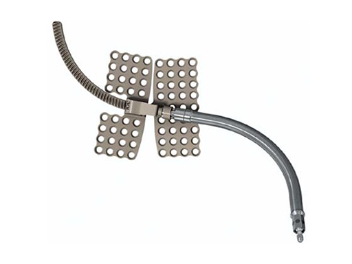
The new 1.3 mm Pediatric Curvilinear Distractor system is an internal distraction osteogenesis device that gradually advances the mandible along a curved trajectory of distraction. As with the previously developed 2.0 mm Curvilinear Distractor, the 1.3 mm is indicated for correction of congenital deficiencies or posttraumatic defects on the mandibular body and ramus, where gradual bone distraction is required. This 1.3 mm pediatric version is intended for patients four years of age and younger.
This pediatric system is part of the curvilinear distractors family, which lengthens the mandible in both vertical and horizontal planes to close an existing open bite, or avoid creating one, secondarily to distraction.
In order to adapt the existing distractor's design to smaller anatomies of younger patients, it was necessary to reduce the overall size of the device in terms of profile (from 7.5 mm to 5.5 mm) and track width (from 4.25 mm to 3 mm). Other modifications include the footplate design, which changed to a mesh pattern to allow for the insert of more screws in a smaller area of bone, and the distractor acceptance of 1.3 mm diameter bone screws.
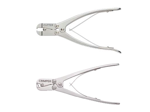
The device allows advancement of up to 35 mm, leaving the surgeon the option of cutting and crimping the track if less advancement is required. Existing tools, such as cutter and crimper were slightly modified to be able to work with both sized distractors: 2.0 mm and 1.3 mm.
A functional stop is created by a track crimp to avoid undesired device disassembling. The distractor is made of titanium molybdenum, is for single use only, and has left and right assemblies available in different radii of curvature (30 mm, 40 mm, 50 mm, 70 mm, and 100 mm), as well as a straight version.
All distractors accept removable extension arms, which move the point of activation to a location that is easily accessible with the activation instrument.
One full rotation of the activation instrument equals 1.0 mm of distraction per day (one half turn twice daily), which is recommended to prevent premature consolidation, and in young patients a rate of 1.5 to 2.0 mm per day could be considered.
Multiplanar mandibular with curvilinear distractor in a 16-month old girl with Treacher Collins syndrome to improve mandibular and airway morphology and achieve tracheostomy removal.
Fig 1ac Proposed curvilinear surgical plan.
Fig 2 ac Lateral facial x-rays. Left to right: Immediately after distractor placement; During distraction; At completion of distraction.
Fig 3ab Pre- and postdistraction 3-D CT scans demonstrating curvilinear lengthening of the mandible.
Fig 4 Predistraction.
Fig 5 During distraction.
Fig 6 Postdistraction. Note significant improvement in mandibular position with closure of open bite deformity.
Fig 7ac Lateral and frontal views 6 months after removal of the device.
Hazards and labeling
Due to varying countries’ legal and regulatory approval requirements, consult the appropriate local product labeling for approved intended use of the products described on this website. All devices on this website are approved by the AO Technical Commission. For logistical reasons, these devices may not be available in all countries worldwide at the date of publication.
Legal restrictions
This work was produced by AO Foundation, Switzerland. All rights reserved by AO Foundation. This publication, including all parts thereof, is legally protected by copyright.
Any use, exploitation or commercialization outside the narrow limits set forth by copyright legislation and the restrictions on use laid out below, without the publisher‘s consent, is illegal and liable to prosecution. This applies in particular to photostat reproduction, copying, scanning or duplication of any kind, translation, preparation of microfilms, electronic data processing, and storage such as making this publication available on Intranet or Internet.
Some of the products, names, instruments, treatments, logos, designs, etc referred to in this publication are also protected by patents, trademarks or by other intellectual property protection laws (eg, “AO” and the AO logo are subject to trademark applications/registrations) even though specific reference to this fact is not always made in the text. Therefore, the appearance of a name, instrument, etc without designation as proprietary is not to be construed as a representation by the publisher that it is in the public domain.
Restrictions on use: The rightful owner of an authorized copy of this work may use it for educational and research purposes only. Single images or illustrations may be copied for research or educational purposes only. The images or illustrations may not be altered in any way and need to carry the following statement of origin “Copyright by AO Foundation, Switzerland”.
Check www.aofoundation.org/disclaimer for more information.
If you have any comments or questions on the articles or the new devices, please do not hesitate to contact us.
“approved by AO Technical Commission” and “approved by AO”
The brands and labels “approved by AO Technical Commission” and “approved by AO”, particularly "AO" and the AO logo, are AO Foundation's intellectual property and subject to trademark applications and registrations, respectively. The use of these brands and labels is regulated by licensing agreements between AO Foundation and the producers of innovation products obliged to use such labels to declare the products as AO Technical Commission or AO Foundation approved solutions. Any unauthorized or inadequate use of these trademarks may be subject to legal action.
AO ITC Innovations Magazine
Find all issues of the AO ITC Innovations Magazine for download here.
Innovation Awards
Recognizing outstanding achievements in development and fostering excellence in surgical innovation.


