Tibia Plateau Leveling Osteotomy Plate 3.5 System (TPLO)
Cranial cruciate ligament (CrCL) injury is the most common cause of hind-limb lameness in the dog. This injury is frequently treated with the tibial plateau leveling osteotomy (TPLO). The TPLO procedure dynamically stabilizes the knee by eliminating cranial tibial subluxation during the weight-bearing phase of locomotion. The slope of the dogs tibial plateau is much greater than its human counterpart, which results in increased loads on the canine CrCL. Unlike ACL injury in humans, many canine CrCL tears start as a partial tear and progress to a complete tear due to degenerative changes occurring in the ligament.
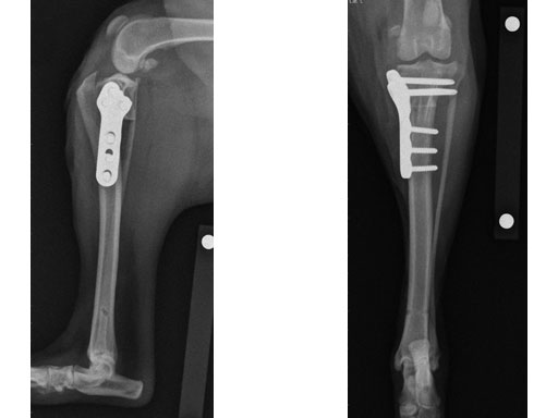
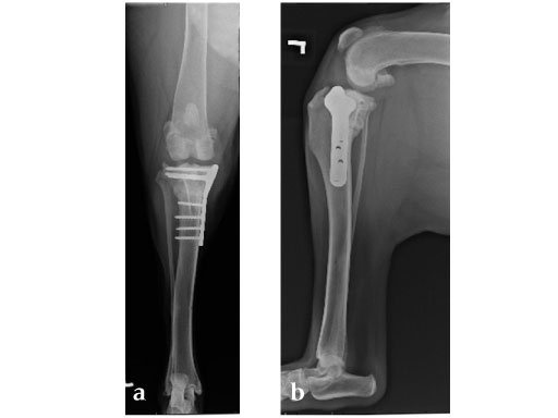
The TPLO system is indicated for osteotomies of the canine proximal tibia and combines plates with a basic instrumentation set. The plates are precontoured to match the anatomic configuration of the medial aspect of the proximal tibia with a limited-contact design and optimal screw placement in the proximal region of the tibia. The plates are available in left and right configurations and feature locking-screw technology.
TPLO is used to treat complete and partial CrCL tears. Early treatment of partial CrCL tears by TPLO has been shown to protect the remaining fibers of the CrCL and reduce the chance of future osteoarthritis and meniscal injury. The osteotomy typically is healed within 8 weeks and return to function can begin progressively after evidence of x-ray healing. The long-term prognosis is excellent.
Until recently, the plates were available in three different profiles: 2.7, 3.5, and 3.5 mm broad. Now, a 3.5 mm plate for small stature was added to the system for use in heavy, but small-boned dogs (ie, bull terriers, bulldogs). The plates and screws are available in stainless steel. The TPLO plates can accommodate locking or cortical screws that correspond to the size of the plate used.
The TPLO 3.5 mm small stature is placed on the medial surface of the tibia. The plate fits very proximally and just distal to the articular surface. In addition, the neck of the plate sits directly over the osteotomy cut. The screw size and pattern provides sufficient strength in the candidate canines tibia. The locking holes are appropriately angled so that the screws will not impinge into the joint or the osteotomy when the plate is applied with proper technique. The head of the plate is also designed to accommodate the placement of a positioning jig.
Biomechanical Consequences of the TPLO Procedure
Objective
Tibia plateau leveling osteotomy (TPLO) is a surgical procedure developed for the treatment of cranial cruciate ligament deficient stifles in dogs. TPLO is proposed to decrease cranial tibial thrust by controlled rotation of the tibial plateau. Currently, conventional and angular stable plate fixation can be performed to stabilize the cylindrical osteotomy. This study investigated the biomechanical consequences of the procedure including the effect of using either conventional or locking head screws in the tibial plateau fragment.
Materials/methods
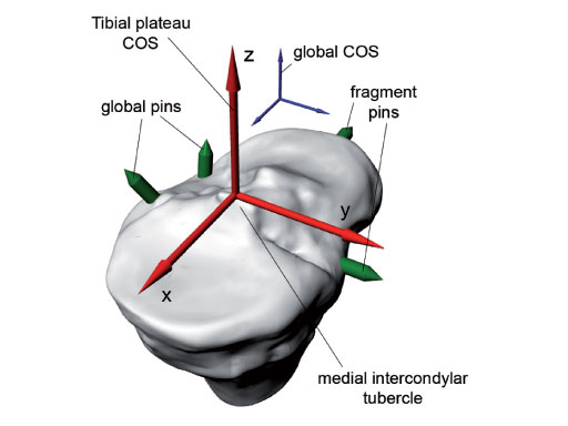
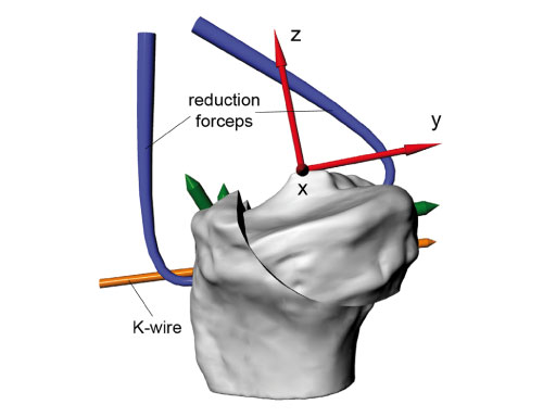
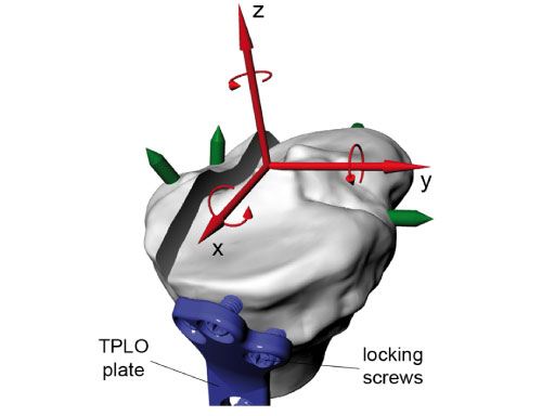
Eight pairs of cadaveric dog tibiae were instrumented with titanium reference pins in order to track the tibial plateau fragment orientation by means of CT imaging. Position of the tibial plateau was determined for the intact bone, after rotation of the fragment, and after plate application (Fig 1). Bones were pairwise instrumented by an experienced surgeon with TPLO plates using either conventional or locking head screws in the tibial plateau fragment. All specimens were biomechanically tested in physiological orientation using cyclic axial compression at 1000 N for 30.000 cycles. Stiffness at the beginning of the test and plastic deformation of the construct at the end of the test were determined in terms of displacement of the machine actuator.
Results
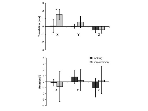
The conventional screw group revealed a significant larger translation of the fragment towards the plate (P =.006) and a greater variance
in fragment rotation as a result of plate application (Fig 2). However, maximum deviations of the achieved tibial plateau orientation from the preoperatively planned orientation were up to 5 for both groups. The amount of this rotation correlated significantly with the plastic deformation of the construct after testing in the conventional screw group (R = 0.81, P = .005). Neither plastic deformation nor construct stiffness were significantly different between conventional and angular stable plate fixation.
Conclusion
This study demonstrated less variation of the tibial plateau position due to instrumentation with an angular stable TPLO plate. However, the fixation type did not affect the biomechanical stability of the construct. Within the conventional group a higher biomechanical stability can be achieved due to a lower degree of rotation which leads to a larger bony contact between the fragments.
With the introduction of the TPLO system, AOVet is also introducing the concept of locked plating to the veterinary practitioners. Therefore, this system also includes a new veterinary basic instrumentation set that contains both standard and locking head screws. The compact design fits into the smaller sized autoclaves commonly used in veterinary clinics.
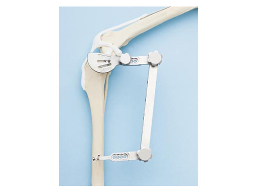
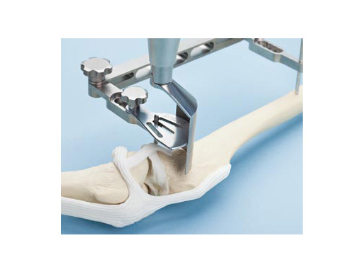
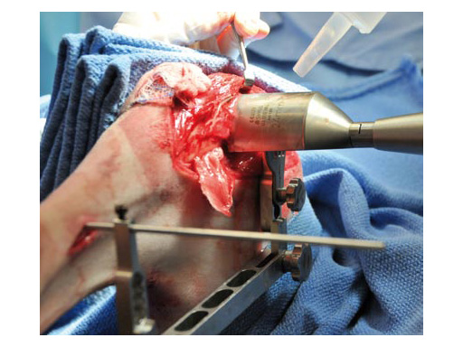
The standard saw and jig are designed specifically for use when performing tibial plateau leveling osteotomy to assist with location, guidance, initiation, and stabilization of the radial saw cut in medium and large breed dogs.
The jig and saw guides can be used bilaterally (for right or left tibiae). Specially designed hinge and saw guide screws within the jig resist loosening under vibration. The hardened steel jig pin screws resist stripping. The saw guides can be positioned in multiple positions along the jig and the jig arm position can be adjusted to allow for optimal placement of the osteotomy. The saw guides are available in three radii of 24 mm, 27 mm, and 30 mm to match the saw blade size selected for an individual patient. The instruments can be easily disassembled for cleaning, and are made of medical grade stainless steel.
A smaller version of the jig and saw guides will be designed in the future for smaller breed dogs.
Case 1: Two-year-old Labrador Retriever
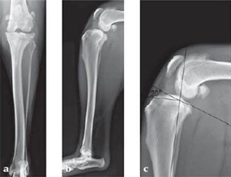
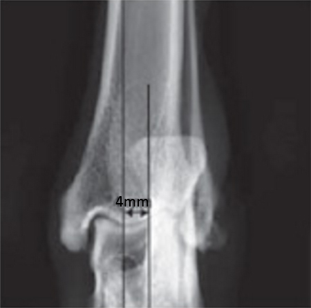
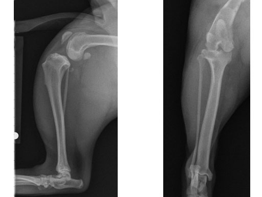
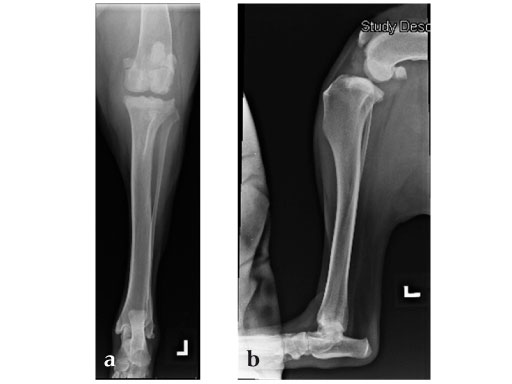
2-years-old Labrador Retriever, 30 kg, female. Chronic lameness in both hind limbs, chronic bilateral cranial cruciate ligament tears, with subsequent stifle joint instability and degenerative joint disease. At that time, she was more clinically lame on the left hind limb, and a surgical correction was subsequently performed on this limb. X-rays of the stifle joint revealed the degenerative joint changes and an effusion; the tibial plateau slope was 20. In addition, the x-rays confirmed that there was a slight amount of tibial torsion that also was observed clinically, accounting for a slight internal rotation of the distal limb. Radiographically, this could be assessed by a 4 mm shift of the normal point of intersection of the medial aspect of the calcaneus with the deepest point of the talar sulcus.
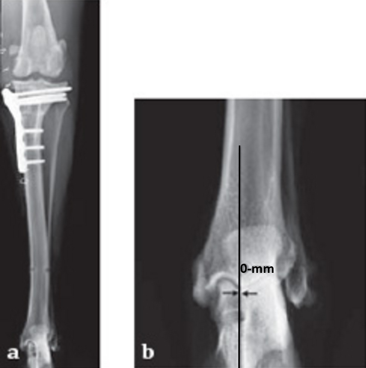
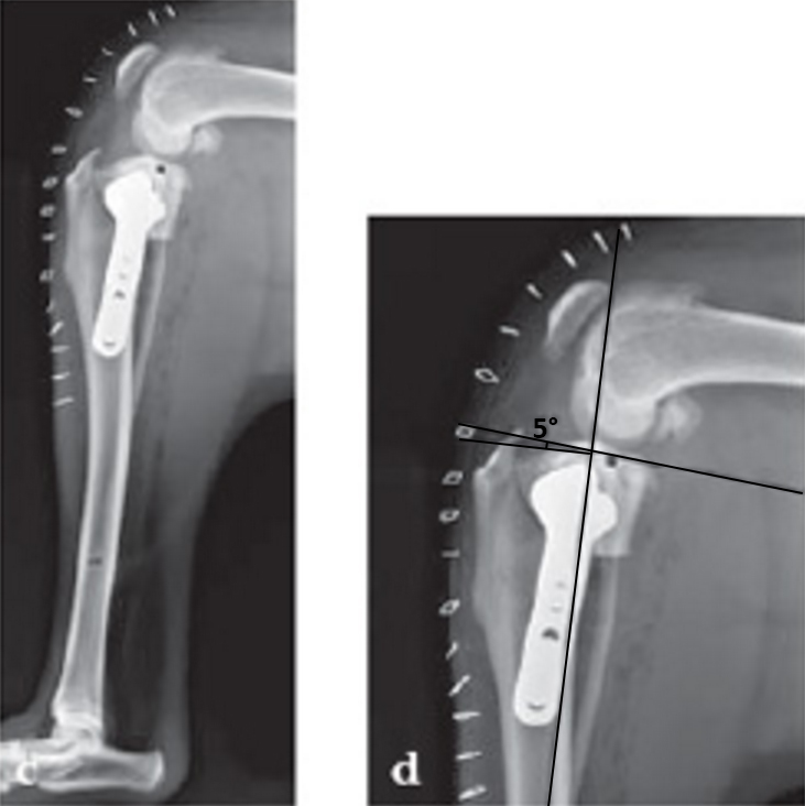
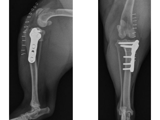
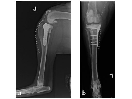
The stifle joint was surgically explored. All remaining remnants of the torn cranial cruciate ligament were debrided; in addition, the caudal pole of the medial meniscus was torn/crushed, and a partial meniscectomy of the damaged portion was performed. A TPLO plate 3.5 was applied to stabilize the fracture. The plate was applied in a neutral fashion. Postoperative x-rays revealed a tibial plateau angle of 5, and a correction of the torsion to 0 mm.
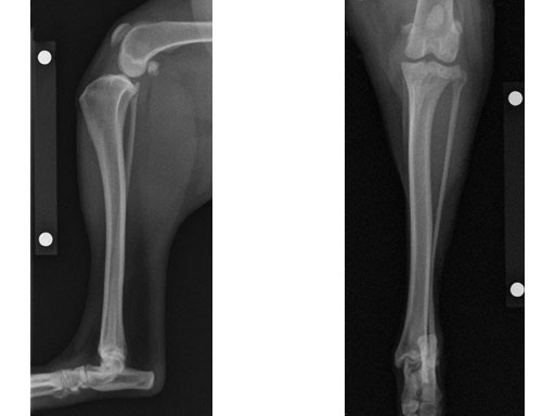
Follow-up x-rays at 8 weeks postoperatively revealed that the osteotomy had healed, and the dog was doing very well. The identical procedure was performed on the opposite stifle joint 2 months later. Healing was again obtained 8 weeks postoperative. Presently, the dog is about 1 year postoperatively and functioning very well.
Case 2: Four-year-old English bulldog
(Case provided by Brian Beale, Houston, USA)
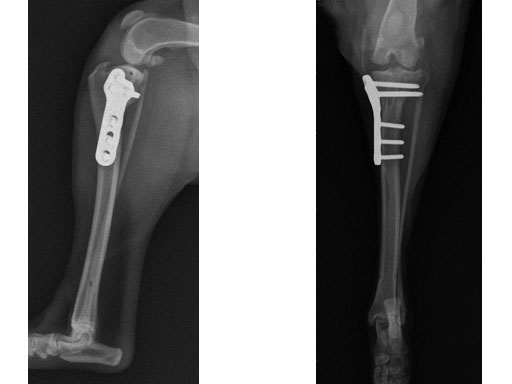
A four-year-old, female, spayed, 33 kg English bulldog had a CrCL tear and a medial patellar luxation. The small stature 3.5 mm TPLO plate was perfect for this dog due to the small profile of the bone and the need to use a heavier plate (3.5 vs a 2.7 mm). In the past, veterinary surgeons have been forced to either squeeze the standard TPLO 3.5 mm plate on the bone or use an undersized TPLO 2.7 mm plate. In this patient, the shorter and smaller profile head of the small stature TPLO 3.5 mm was perfect.
Case 3: Eight-year-old Australian cattle dog
(Case provided by Brian Beale, Houston, USA)
An 8-year-old, female, spayed, 24 kg Australian cattle dog. This breed has short stocky legs and is very energetic and strong. The added strength of the 3.5 mm plate over the TPLO 2.7 mm plate was an advantage. The smaller head profile and shorter length of the TPLO 3.5 mm small stature plate allowed it to fit nicely on this patient.
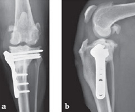
Case 4: Three-year-old neutered female Tosa, 60 kg, chronic (6 months) lameness, painful stifle.
(Case provided by Randy Boudrieau, North Grafton, USA)
Hazards and labeling
Due to varying countries’ legal and regulatory approval requirements, consult the appropriate local product labeling for approved intended use of the products described on this website. All devices on this website are approved by the AO Technical Commission. For logistical reasons, these devices may not be available in all countries worldwide at the date of publication.
Legal restrictions
This work was produced by AO Foundation, Switzerland. All rights reserved by AO Foundation. This publication, including all parts thereof, is legally protected by copyright.
Any use, exploitation or commercialization outside the narrow limits set forth by copyright legislation and the restrictions on use laid out below, without the publisher‘s consent, is illegal and liable to prosecution. This applies in particular to photostat reproduction, copying, scanning or duplication of any kind, translation, preparation of microfilms, electronic data processing, and storage such as making this publication available on Intranet or Internet.
Some of the products, names, instruments, treatments, logos, designs, etc referred to in this publication are also protected by patents, trademarks or by other intellectual property protection laws (eg, “AO” and the AO logo are subject to trademark applications/registrations) even though specific reference to this fact is not always made in the text. Therefore, the appearance of a name, instrument, etc without designation as proprietary is not to be construed as a representation by the publisher that it is in the public domain.
Restrictions on use: The rightful owner of an authorized copy of this work may use it for educational and research purposes only. Single images or illustrations may be copied for research or educational purposes only. The images or illustrations may not be altered in any way and need to carry the following statement of origin “Copyright by AO Foundation, Switzerland”.
Check www.aofoundation.org/disclaimer for more information.
If you have any comments or questions on the articles or the new devices, please do not hesitate to contact us.
“approved by AO Technical Commission” and “approved by AO”
The brands and labels “approved by AO Technical Commission” and “approved by AO”, particularly "AO" and the AO logo, are AO Foundation's intellectual property and subject to trademark applications and registrations, respectively. The use of these brands and labels is regulated by licensing agreements between AO Foundation and the producers of innovation products obliged to use such labels to declare the products as AO Technical Commission or AO Foundation approved solutions. Any unauthorized or inadequate use of these trademarks may be subject to legal action.
AO ITC Innovations Magazine
Find all issues of the AO ITC Innovations Magazine for download here.
Innovation Awards
Recognizing outstanding achievements in development and fostering excellence in surgical innovation.


