Double/Triple Pelvic Osteotomy (DPO/TPO) plate
Erik Asimus, Brian Beale, Randy J Boudrieau, Loïc Déjardin, Michael Kowaleski
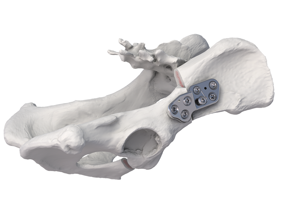
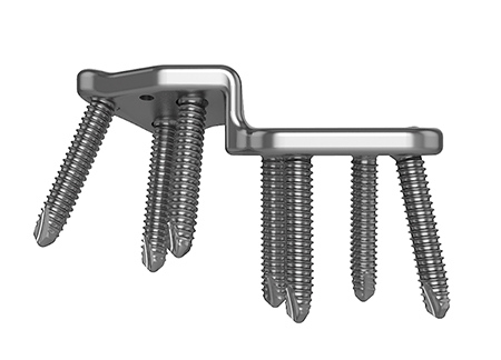
The DePuy Synthes Vet double/triple pelvic osteotomy (DPO/TPO) plate is indicated for treating coxofemoral joint instability and subluxation in immature dogs prior to the onset of osteoarthritis. The DPO/TPO plate is 3.2 mm thick and available in right and left versions with angulations of 20°, 25° and 30° between the plate surfaces so as to perform the rotational osteotomy of the acetabular bone segment.
Background
At least 3.5% of the global dog population suffers from hip dysplasia [1]. In fact, the incidence of hip dysplasia can be up to 50% in some larger breeds of dogs [2]. Rotational pelvic osteotomies are prophylactic surgical interventions intended to decrease abnormal hip joint laxity, normalize articular stresses and improve hip joint congruity. Currently, the DPO and TPO are the most popular corrective procedures. However, despite technique modifications and development of new plates, complications such as implant loosening, reduction of the pelvic inlet diameter, over- or under-rotation of the acetabular rim and delayed healing of the osteotomies can occur.
Plate Design
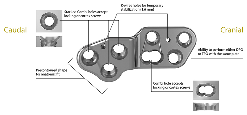
The recently launched DPO/TPO plate offers substantial improvements to existing bone fixation plates to overcome these complications (Fig 2). Features of the plate include:
- Screw trajectories designed to optimize screw purchase into the relatively soft bone.
- Anatomically contoured to match the ilial shaft and to allow clearance for acetabular flare and the tuberosity at the origin of the rectus femoris muscle.
- Plate design includes two distinct screw-hole technologies accommodate all plating modalities (stacked combi holes and coaxial combi-hole).
- Incorporation of locking technology permits a fixed-angle device to increase construct strength.
References
1 LaFond E, Breur GJ, Austin CC. Breed susceptibility for developmental orthopedic diseases in dogs. J Am Anim Hosp Assoc. 2002 SepOct; 38(5):46777.
2 Todhunter RJ, Mateescu R, Lust G, et al. Quantitative trait loci for hip dysplasia in a cross-breed canine pedigree. Mamm Genome. 2005 Sep; 16(9):72030.
Case 1: Labrador retriever puppy
(Case provided by Brian Beale, Houston, USA)
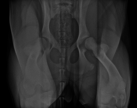
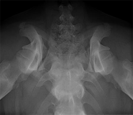
A 5-month-old spayed female labrador retriever puppy weighing 22.0 kg presented with bilateral hind limb weakness and a bunny-hopping gait in the hind limbs. Physical examination revealed bilateral hip instability (positive Ortolani sign) and mild pain on full extension of the hips. Slight crepitus was palpated in the left hip. The gluteal muscles appeared to have mild atrophy. The neurological exam was normal. Radiographic examinations revealed bilateral hip subluxation and a distraction index of 0.5 of the right hip and 0.7 of the left hip (Fig 3). No evidence of osteoarthritis was observed.
A diagnosis of juvenile hip dysplasia was made. The right hip was considered an acceptable candidate for double pelvic osteotomy (DPO; note that in a 5-month-old dog, an osteotomy of the pubis (as performed in a TPO) is not necessary due to the bony compliance at this young age). The left hip conformation was considered too abnormal for a corrective osteotomy and was not treated. The owner was counseled that a total hip replacement (THR) may be needed in the future on the left hip.
Angles of subluxation (10°) and reduction (30°) of the right hip were measured under anesthesia and the patient was placed in dorsal recumbency. A 7 mm portion of the right pubic body was excised. The patient was repositioned into lateral recumbency. A right ilial osteotomy was made immediately caudal to the sacrum. A 25° DPO/TPO plate was attached to the caudal ilial bone segment using locking 3.5 mm screws in the three stacked combi holes. The caudal acetabular segment was rotated laterally until the cranial aspect of the plate was in contact with the lateral aspect of the cranial ilial segment. The osteotomy site was compressed, and the plate was secured to the cranial ilial bone segment using a 3.5 mm cortical screw in the LCP combi hole in the cranial side of the plate. Three additional 3.5 mm locking screws were placed in the remaining stacked combi holes in the cranial segment of the plate.
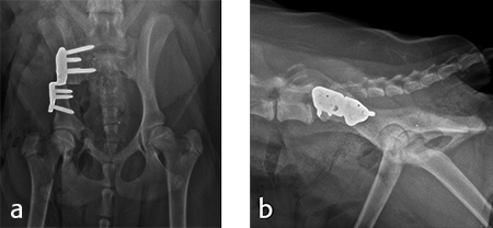
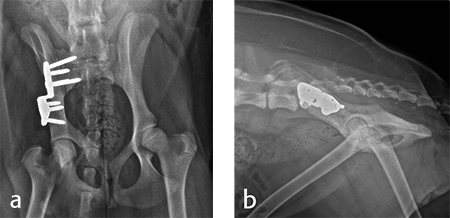
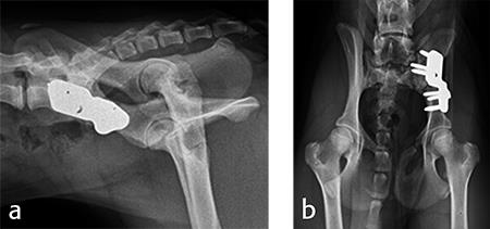
Postoperative radiographs revealed reduction in subluxation with capture of the femoral head in the right coxofemoral joint (Fig 4). Palpation of the hip revealed good stability of the right hip. Activity was restricted to leash walk only for 6 weeks postoperatively. Radiographic examination 7 weeks following surgery revealed healing of the ilial osteotomy, stable implants, and excellent coxofemoral conformation and stability (Fig 5).
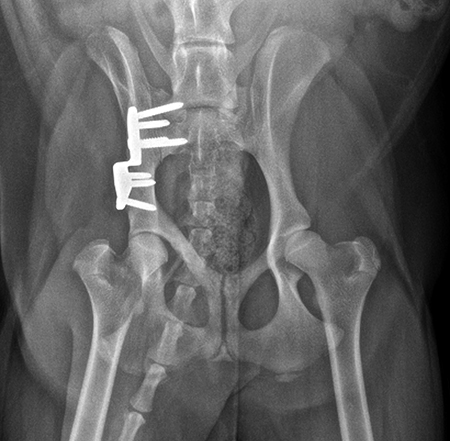
Radiographic examination at 6 months postsurgery revealed stable implants, excellent coxofemoral conformation, and no evidence of osteoarthritis of the right hip. The left acetabulum was mildly shallow and mild subluxation of the femoral head was present at follow-up examination (Fig 6). Early osteophytosis in the region of the left femoral neck was evident. The dog was using the right hind leg normally and was showing no signs of instability or pain of the right hip. Mild instability and pain of the left hip was present on palpation. The dogs left hip was treated with a joint supplement and NSAIDs as needed. Future THR will be performed if clinical signs no longer respond to medical treatment.
Case 2: Boxer puppy
(Case provided by Erik Asimus, Toulouse, France)
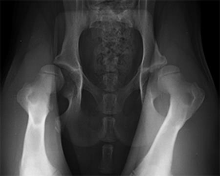
A 4-month-old female boxer puppy weighing 15.0 kg presented with bilateral hind limb weakness and reluctance to walk. Physical examination revealed bilateral hip instability (positive Ortolani sign) and severe pain on full extension of the hips. The neurological exam was normal. The radiographs revealed bilateral hip subluxation and a distraction index of 0.65 of the right hip and 0.6 of the left hip (Fig 7). Very mild osteoarthritis was seen and femoral head coverage by the dorsal acetabular rim was good (Fig 8). Angles of subluxation (10° R and 20° L) and reduction (30° R and 40° L) of the hips were measured under anesthesia.
A diagnosis of juvenile hip dysplasia was made. Both hips were considered as candidate for double pelvic osteotomy (DPO). Considering the difficulty to limit the activity of this active puppy, a simultaneous bilateral procedure was not performed. The left hip DPO was performed first, followed by the right hip 4 weeks later.
For each surgical procedure, the patient was placed in dorsal recumbency to enable the pubic ostectomy. The patient was repositioned in lateral recumbency to perform the DPO. A left ilial osteotomy was performed caudal to the sacrum. A 25° DPO/TPO plate was attached to the caudal ilial segment using locking 3.5 mm screws in the three stacked combi holes. The caudal acetabular segment was rotated laterally until the cranial aspect of the plate was in contact with the lateral aspect of the cranial ilial segment. The osteotomy site was compressed and the plate was secured to the cranial ilial bone segment using a 3.5 mm cortical screw in the LCP combi hole in the cranial side of the plate. Three additional 3.5 mm locking screws were placed in the remaining stacked combi holes in the cranial segment of the plate (Fig 9).
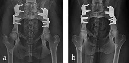
Activity was restricted to leash walks for 6 weeks postoperatively. The radiographic examination 1 month after each surgery revealed partial healing of the ilial osteotomy and stable implants. Postoperative radiographs at 6 months after both surgical procedures revealed complete healing of the ilial osteotomies, stable implants, and excellent coxofemoral conformation, with no subluxation of the femerol head. Mild osteoarthritis was observed, however. At both the 4 and 6 month evaluation, the dog was using both hind limbs without any evidence of lameness and was showing no signs of instability or pain of either hip (Fig 10).
“approved by AO Technical Commission” and “approved by AO”
The brands and labels “approved by AO Technical Commission” and “approved by AO”, particularly "AO" and the AO logo, are AO Foundation's intellectual property and subject to trademark applications and registrations, respectively. The use of these brands and labels is regulated by licensing agreements between AO Foundation and the producers of innovation products obliged to use such labels to declare the products as AO Technical Commission or AO Foundation approved solutions. Any unauthorized or inadequate use of these trademarks may be subject to legal action.
AO ITC Innovations Magazine
Find all issues of the AO ITC Innovations Magazine for download here.
Innovation Awards
Recognizing outstanding achievements in development and fostering excellence in surgical innovation.


