LCP Meta-Diaphyseal Volar Distal Radius Plate
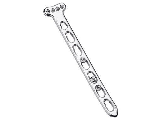
The LCP meta-diaphyseal volar distal radius plates have been designed as longer distal radius plates, to treat fractures with a proximal extension into the diaphyseal regions of the radius.
The meta-diaphyseal volar distal radius plate is effectively a plate combining the head of the LCP volar distal radius plate with a similar shaft to the LCP 3.5. But the meta-diaphyseal volar distal radius plate can be placed more proximal than the LCP volar distal radius plate, and it is significantly stronger.
The meta-diaphyseal volar distal radius plate is precontoured to match the volar surface of the distal radius. It features a 25 angulation equivalent to the normal anatomy of the distal radius. The plate head offers 4 threaded round holes which accept 2.4?mm locking head screws. The plates are available with 5, 7, 9, 11, 13, or 15 holes in the shaft, ranging from 95240?mm. A precontoured radial bow is included in the shaft of the plates after 95?mm to match the anatomic curve of the radial shaft. Relief cuts are included after every other hole, beginning after the 7th hole (after 95?mm) to ease additional contouring needed to match the patients specific radial bow. The plates are available in left and right configurations and in stainless steel or CP titanium.
Technical Know-how in Image Processing
We support your Clinical Development and Publication
About us
The Human Morphology Service Center (HMSC) of the AO Development Institute (ADI) maintains a database of CT scans of cadaveric human specimens used in other projects in the AO institutes. Additionally, the database includes virtual bone models of the entire skeleton which are derived from the CT scans. This database is registered at the relevant regulatory authorities in Switzerland. A major task of the HMSC is to offer support in implant design and optimization to the AO Network and external partners, based on its database, know-how, and resources.
Services
Since its foundation in 2002 the HMSC has acquired more than a thousand bone data sets, know-how in medical image computing, and statistical shape analysis of 3-D bone models. Moreover, it has excellent software resources (Amira, Geomagic, Unigraphics, Matlab) to accomplish its support task. The service offerings include CT scans of cadaveric bones, image processing, segmentation, reverse engineering, statistical shape analysis, principal components analysis (PCA), rapid prototyping and CMF related image computing.
Example
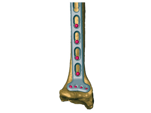
An example of our services is the response to a request from the AO Hand Expert Group (HAEG) which was interested in the shape variability of the distal radius and shaft, with respect to the meta-diaphyseal volar distal radius plate. Together with a medical and a technical mentor, a sample of 15 radii from the database was selected and evaluated. To identify the region of interest (ROI) on the virtual specimens, we aligned a virtual template of a plate prototype on the radii, and placed landmarks on the bone surfaces through the first eight distal plate holes as illustrated in Fig 1. These landmarks were used to align the bones to each other by a generalized Procrustes fit, which minimises the sum of the distances between homologous landmarks.
Fig 1: The placement of homologous landmarks on the radius surface through the holes of a manually aligned template of a meta-diaphyseal volar distal radius plate prototype.
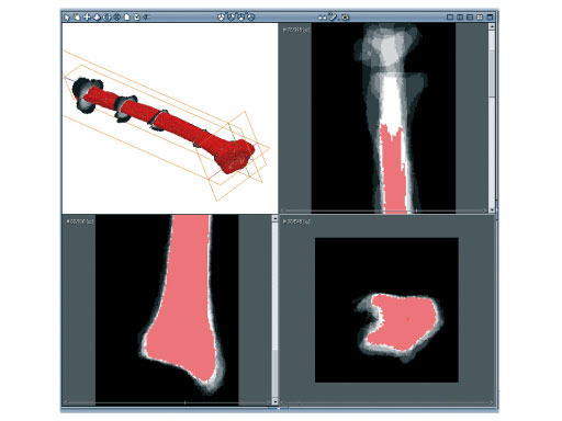
Then, the aligned surfaces were scan-converted and superposed, resulting in a 3-D image in which the grey values of the voxels represent the number of bone model intersections as illustrated in Fig 2. This 3-D image carries statistical information for the complete sample. It enables us to extract the median surface and to visualize local deviations from the median bone.
Fig 2: The different cuts through the superposed radii give insight into the shape variability with respect to the region of interest (the plate). The red color represents the median segmentation used for median surface generation.
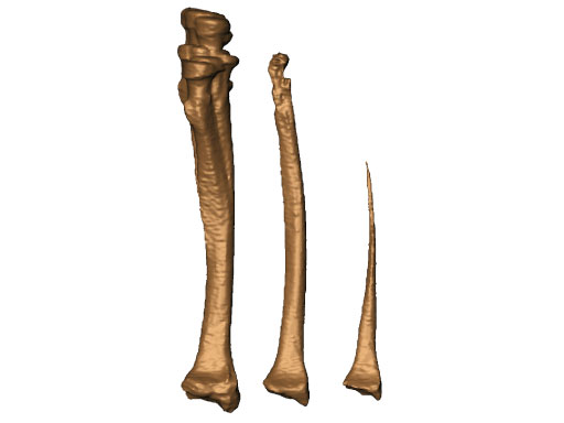
Fig 3 shows three surfaces of the aligned and superposed volumes of the radii:
1. envelope of all radii
2. median intersection
3. common intersection
The median region of interest (ROI) is intended to be a template for potential preshaped plates. It can be observed that the shape variability is very small around the landmarks used for aligning the bones and that the variability increases with a growing distance from them. By carefully choosing only the ROI relevant for the implant, the aligning and averaging of the bone surface results in optimal templates for preshaped implants. The implant has to fit well only to the ROI. Variations outside are of no interest for the plate design. Such investigations can result in a basis for decision-making to engineers of implant manufacturers.
Fig 3: The surfaces of the envelope, the median, and the common intersection surfaces (from left to right) of the aligned and superposed volumes of the radii of the sample.
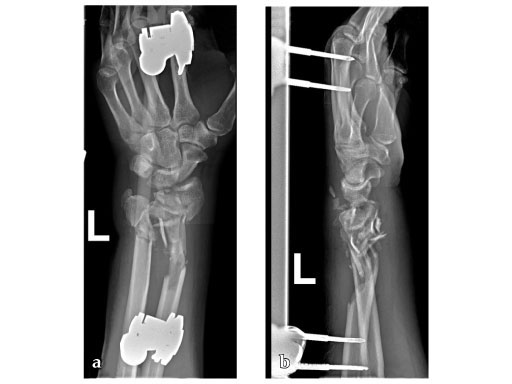
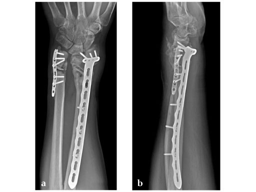
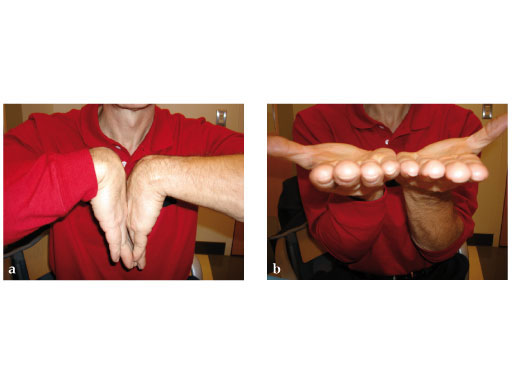
34-year-old male, with open fracture after motor vehicle accident.
Fig 1ab: Preoperative x-rays; primary stabilization with external fixator.
Fig 2ab: Eight months postoperative; full forearm rotation and 75% grip strength and wrist motion.
Fig 3ab: Full motion recovery.
Case provided by Jesse Jupiter, Boston
Hazards and labeling
Due to varying countries’ legal and regulatory approval requirements, consult the appropriate local product labeling for approved intended use of the products described on this website. All devices on this website are approved by the AO Technical Commission. For logistical reasons, these devices may not be available in all countries worldwide at the date of publication.
Legal restrictions
This work was produced by AO Foundation, Switzerland. All rights reserved by AO Foundation. This publication, including all parts thereof, is legally protected by copyright.
Any use, exploitation or commercialization outside the narrow limits set forth by copyright legislation and the restrictions on use laid out below, without the publisher‘s consent, is illegal and liable to prosecution. This applies in particular to photostat reproduction, copying, scanning or duplication of any kind, translation, preparation of microfilms, electronic data processing, and storage such as making this publication available on Intranet or Internet.
Some of the products, names, instruments, treatments, logos, designs, etc referred to in this publication are also protected by patents, trademarks or by other intellectual property protection laws (eg, “AO” and the AO logo are subject to trademark applications/registrations) even though specific reference to this fact is not always made in the text. Therefore, the appearance of a name, instrument, etc without designation as proprietary is not to be construed as a representation by the publisher that it is in the public domain.
Restrictions on use: The rightful owner of an authorized copy of this work may use it for educational and research purposes only. Single images or illustrations may be copied for research or educational purposes only. The images or illustrations may not be altered in any way and need to carry the following statement of origin “Copyright by AO Foundation, Switzerland”.
Check www.aofoundation.org/disclaimer for more information.
If you have any comments or questions on the articles or the new devices, please do not hesitate to contact us.
“approved by AO Technical Commission” and “approved by AO”
The brands and labels “approved by AO Technical Commission” and “approved by AO”, particularly "AO" and the AO logo, are AO Foundation's intellectual property and subject to trademark applications and registrations, respectively. The use of these brands and labels is regulated by licensing agreements between AO Foundation and the producers of innovation products obliged to use such labels to declare the products as AO Technical Commission or AO Foundation approved solutions. Any unauthorized or inadequate use of these trademarks may be subject to legal action.
AO ITC Innovations Magazine
Find all issues of the AO ITC Innovations Magazine for download here.
Innovation Awards
Recognizing outstanding achievements in development and fostering excellence in surgical innovation.


